PI3 Kinase, class II a (PI3KC2A) is also known as phosphatidylinositol-4-phosphate 3-kinase C2 domain-containing subunit alpha. It plays an important role in vesicular transport in eukaryotic cells. PI3 Kinase-C2-alpha also plays a part in cellular proliferation and survival.
Product Information
Format
Purified
Control
Jurkat cell lysate
Presentation
Purified mouse monoclonal IgMκ in buffer containing PBS with 0.05% sodium azide.
Applications
Application
Anti-PI3 Kinase-C2-alpha Antibody, clone 9H4.2 detects level of PI3 Kinase-C2-alpha & has been published & validated for use in WB & IC.
Key Applications
Western Blotting
Immunocytochemistry
Application Notes
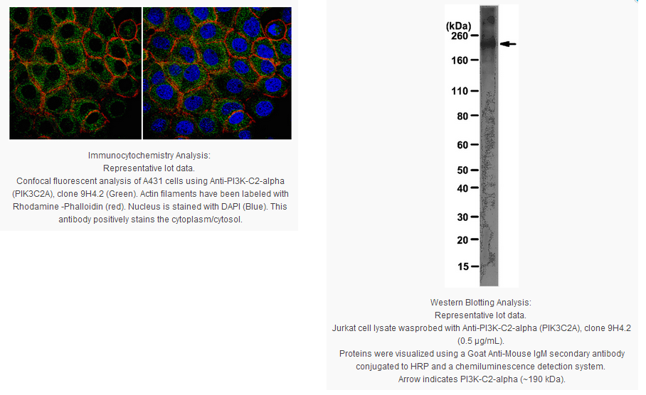
Immunocytochemistry Analysis: 1:500 dilution from a previous lot detected PI3K-C2-alpha in A431 cells.
Biological Information
Immunogen
KLH-conjugated linear peptide corresponding to human PI3K-C2-alpha.
Clone
9H4.2
Concentration
Please refer to the Certificate of Analysis for the lot-specific concentration.
The protein encoded by this gene belongs to the phosphoinositide 3-kinase (PI3K) family. PI3-kinases play roles in signaling pathways involved in cell proliferation, oncogenic transformation, cell survival, cell migration, and intracellular protein trafficking. This protein contains a lipid kinase catalytic domain as well as a C-terminal C2 domain, a characteristic of class II PI3-kinases. C2 domains act as calcium-dependent phospholipid binding motifs that mediate translocation of proteins to membranes, and may also mediate protein-protein interactions. The PI3-kinase activity of this protein is not sensitive to nanomolar levels of the inhibitor wortmanin. This protein was shown to be able to be activated by insulin and may be involved in integrin-dependent signaling. [provided by RefSeq].
FUNCTION: Phosphorylates PtdIns, PtdIns4P and PtdIns(4,5)P2. May play a role in clathrin-coated endocytic vesicle formation and EGF signaling cascade. May be involved in mitosis and UV-induced damage response. May be a downstream effector in insulin signaling cascade.
COFACTOR: Calcium or magnesium. Manganese cannot be used.
ENZYME REGULATION: Activated by insulin By similarity. Only slightly inhibited by wortmannin and LY294002. Activated by clathrin.
SUBUNIT STRUCTURE: Part of a complex with ERBB2 and EGFR. Interacts with clathrin trimers.
SUBCELLULAR LOCATION: Cell membrane. Golgi apparatus. Cytoplasmic vesicle › clathrin-coated vesicle. Nucleus. Cytoplasm. Note: According to Ref.5 and Ref.8, it is found in the cell membrane, the Golgi apparatus and in clathrin-coated vesicles. According to Ref.18 it inserts preferentially into membranes containing PtdIns(4,5)P2. According to Ref.7, it is nuclear and cytoplasmic. Associated with RNA-containing structures. According to Ref.9, it is mainly cytoplasmic.
TISSUE SPECIFICTY: Expressed in columnar and transitional epithelia, mononuclear cells, smooth muscle cells, and endothelial cells lining capillaries and small venules (at protein level). Ubiquitously expressed, with highest levels in heart, placenta and ovary, and lowest levels in the kidney.
PTM: Phosphorylated upon insulin stimulation; which may lead to enzyme activation (By similarity). Phosphorylated on Ser-259 during mitosis and upon UV irradiation; which does not change enzymatic activity but leads to proteasomal degradation. Ser-259 phosphorylation may be mediated by CDK1 or JNK, depending on the physiological state of the cell.
SEQUENCE SIMILARITIES: Belongs to the PI3/PI4-kinase family.
In the absence of the carrier phosphatidylserine, enzymatic kinetics toward PtdIns4P are non-linear.
KM=122 µM for PtdIns (in the absence of phosphatidylserine) Ref.1
KM=64 µM for PtdIns (in the presence of phosphatidylserine)
KM=25 µM for PtdIns4P (in the presence of phosphatidylserine)
KM=15 µM for ATP (with PtdIns as substrate) (in the absence of phosphatidylserine)
KM=32 µM for ATP (with PtdIns as substrate) (in the presence of phosphatidylserine)
KM=54 µM for ATP (with PtdIns4P as substrate) (in the presence of phosphatidylserine)
Vmax=990 pmol/min/mg enzyme with PtdIns as substrate (in the absence of phosphatidylserine)
Vmax=200 pmol/min/mg enzyme with PtdIns as substrate (in the presence of phosphatidylserine)
Vmax=240 pmol/min/mg enzyme with PtdIns4P as substrate (in the presence of phosphatidylserine)
Vmax=6800 pmol/min/mg enzyme toward ATP with PtdIns as substrate (in the absence of phosphatidylserine)
Vmax=805 pmol/min/mg enzyme toward ATP with PtdIns as substrate (in the presence of phosphatidylserine)
Vmax=880 pmol/min/mg enzyme toward ATP with PtdIns4P as substrate (in the presence of phosphatidylserine)
Product Usage Statements
Quality Assurance
Evaluated by Western Blot in Jurkat cell lysate.
Western Blot Analysis: 0.5 µg/mL of this antibody detected PI3K-C2-alpha on 10 µg of Jurkat cell lysate.
Usage Statement
Unless otherwise stated in our catalog or other company documentation accompanying the product(s), our products are intended for research use only and are not to be used for any other purpose, which includes but is not limited to, unauthorized commercial uses, in vitro diagnostic uses, ex vivo or in vivo therapeutic uses or any type of consumption or application to humans or animals.
Storage and Shipping Information
Storage Conditions
Stable for 1 year at 2-8°C from date of receipt.
Packaging Information
Material Size
100 µg
原厂资料:
Key Spec Table
Species Reactivity
Key Applications
Host
Format
Antibody Type
M, H, R
WB, ICC
M
Purified
Monoclonal Antibody
Description
Catalogue Number
05-1560
Description
Anti-PI3 Kinase-C2-alpha Antibody, clone 9H4.2
Alternate Names
phosphoinositide-3-kinase, class 2, alpha polypeptide
PI3 Kinase, class II a (PI3KC2A) is also known as phosphatidylinositol-4-phosphate 3-kinase C2 domain-containing subunit alpha. It plays an important role in vesicular transport in eukaryotic cells. PI3 Kinase-C2-alpha also plays a part in cellular proliferation and survival.
Product Information
Format
Purified
Control
Jurkat cell lysate
Presentation
Purified mouse monoclonal IgMκ in buffer containing PBS with 0.05% sodium azide.
Applications
Application
Anti-PI3 Kinase-C2-alpha Antibody, clone 9H4.2 detects level of PI3 Kinase-C2-alpha & has been published & validated for use in WB & IC.
Key Applications
Western Blotting
Immunocytochemistry
Application Notes
Immunocytochemistry Analysis: 1:500 dilution from a previous lot detected PI3K-C2-alpha in A431 cells.
Biological Information
Immunogen
KLH-conjugated linear peptide corresponding to human PI3K-C2-alpha.
Clone
9H4.2
Concentration
Please refer to the Certificate of Analysis for the lot-specific concentration.
The protein encoded by this gene belongs to the phosphoinositide 3-kinase (PI3K) family. PI3-kinases play roles in signaling pathways involved in cell proliferation, oncogenic transformation, cell survival, cell migration, and intracellular protein trafficking. This protein contains a lipid kinase catalytic domain as well as a C-terminal C2 domain, a characteristic of class II PI3-kinases. C2 domains act as calcium-dependent phospholipid binding motifs that mediate translocation of proteins to membranes, and may also mediate protein-protein interactions. The PI3-kinase activity of this protein is not sensitive to nanomolar levels of the inhibitor wortmanin. This protein was shown to be able to be activated by insulin and may be involved in integrin-dependent signaling. [provided by RefSeq].
FUNCTION: Phosphorylates PtdIns, PtdIns4P and PtdIns(4,5)P2. May play a role in clathrin-coated endocytic vesicle formation and EGF signaling cascade. May be involved in mitosis and UV-induced damage response. May be a downstream effector in insulin signaling cascade.
COFACTOR: Calcium or magnesium. Manganese cannot be used.
ENZYME REGULATION: Activated by insulin By similarity. Only slightly inhibited by wortmannin and LY294002. Activated by clathrin.
SUBUNIT STRUCTURE: Part of a complex with ERBB2 and EGFR. Interacts with clathrin trimers.
SUBCELLULAR LOCATION: Cell membrane. Golgi apparatus. Cytoplasmic vesicle › clathrin-coated vesicle. Nucleus. Cytoplasm. Note: According to Ref.5 and Ref.8, it is found in the cell membrane, the Golgi apparatus and in clathrin-coated vesicles. According to Ref.18 it inserts preferentially into membranes containing PtdIns(4,5)P2. According to Ref.7, it is nuclear and cytoplasmic. Associated with RNA-containing structures. According to Ref.9, it is mainly cytoplasmic.
TISSUE SPECIFICTY: Expressed in columnar and transitional epithelia, mononuclear cells, smooth muscle cells, and endothelial cells lining capillaries and small venules (at protein level). Ubiquitously expressed, with highest levels in heart, placenta and ovary, and lowest levels in the kidney.
PTM: Phosphorylated upon insulin stimulation; which may lead to enzyme activation (By similarity). Phosphorylated on Ser-259 during mitosis and upon UV irradiation; which does not change enzymatic activity but leads to proteasomal degradation. Ser-259 phosphorylation may be mediated by CDK1 or JNK, depending on the physiological state of the cell.
SEQUENCE SIMILARITIES: Belongs to the PI3/PI4-kinase family.
In the absence of the carrier phosphatidylserine, enzymatic kinetics toward PtdIns4P are non-linear.
KM=122 µM for PtdIns (in the absence of phosphatidylserine) Ref.1
KM=64 µM for PtdIns (in the presence of phosphatidylserine)
KM=25 µM for PtdIns4P (in the presence of phosphatidylserine)
KM=15 µM for ATP (with PtdIns as substrate) (in the absence of phosphatidylserine)
KM=32 µM for ATP (with PtdIns as substrate) (in the presence of phosphatidylserine)
KM=54 µM for ATP (with PtdIns4P as substrate) (in the presence of phosphatidylserine)
Vmax=990 pmol/min/mg enzyme with PtdIns as substrate (in the absence of phosphatidylserine)
Vmax=200 pmol/min/mg enzyme with PtdIns as substrate (in the presence of phosphatidylserine)
Vmax=240 pmol/min/mg enzyme with PtdIns4P as substrate (in the presence of phosphatidylserine)
Vmax=6800 pmol/min/mg enzyme toward ATP with PtdIns as substrate (in the absence of phosphatidylserine)
Vmax=805 pmol/min/mg enzyme toward ATP with PtdIns as substrate (in the presence of phosphatidylserine)
Vmax=880 pmol/min/mg enzyme toward ATP with PtdIns4P as substrate (in the presence of phosphatidylserine)
Product Usage Statements
Quality Assurance
Evaluated by Western Blot in Jurkat cell lysate.
Western Blot Analysis: 0.5 µg/mL of this antibody detected PI3K-C2-alpha on 10 µg of Jurkat cell lysate.
Usage Statement
Unless otherwise stated in our catalog or other company documentation accompanying the product(s), our products are intended for research use only and are not to be used for any other purpose, which includes but is not limited to, unauthorized commercial uses, in vitro diagnostic uses, ex vivo or in vivo therapeutic uses or any type of consumption or application to humans or animals.


 京公网安备11010802025653 版权所有:北京逸优科技有限公司
