PHD2 (Prolyl hydroxylase domain-containing protein 2), also known as Hypoxia-inducible factor prolyl hydroxylase 2 (HIF-PH2 or HPH-2), is one of 4 PHD (PHD1-4) proteins which function as prolyl hydroxylases to effect oxygen homeostasis via hypoxia-inducible factor (HIF). This transcriptional complex is composed of an alpha-beta heterodimer that mediates a broad range of cellular and systemic responses to hypoxia. The PHD proteins hydroxylate HIF-1 alpha at 'Pro-402' and 'Pro-564', and HIF-2 alpha. The PHD2 isoform is the main down-regulator of HIFs for normal oxygen levels and mild hypoxia. When oxygen levels are normal, PHDs catalyze the hydroxylation of prolyl residues on HIF-1alpha. This post-translational modification targets this protein for proteasomal degradation via the von Hippel-Lindau ubiquitination complex.
Product Information
Format
Purified
Control
MCF-7 cell lysate
Presentation
Purified mouse monoclonal IgG1κ in buffer containing 0.1 M Tris-Glycine (pH 7.4, 150 mM NaCl) with 0.05% sodium azide.
Applications
Application
Use Anti-PHD2 Antibody, clone 76a (mouse monoclonal antibody) validated in WB to detect PHD2 also known as EGL nine (C.elegans) homolog 1, HIF prolyl hydroxylase 2, Hypoxia-inducible factor prolyl hydroxylase 2.
Key Applications
Western Blotting
Biological Information
Immunogen
Recombinant protein corresponding to human PHD2.
Epitope
Unknown
Clone
76a
Concentration
Please refer to the Certificate of Analysis for the lot-specific concentration.
FUNCTION: Catalyzes the post-translational formation of 4-hydroxyproline in hypoxia-inducible factor (HIF) alpha proteins. Hydroxylates HIF-1 alpha at 'Pro-402' and 'Pro-564', and HIF-2 alpha. Functions as a cellular oxygen sensor and, under normoxic conditions, targets HIF through the hydroxylation for proteasomal degradation via the von Hippel-Lindau ubiquitination complex.
ENZYME REGULATION: Following exposure to hypoxia, activated in HeLa cells but not in cardiovascular cells. Seems to be inhibited by ING4.
SUBUNIT STRUCTURE: Monomer. Interacts with ING4.
TISSUE SPECIFICITY: According to Ref.1 widely expressed with highest levels in skeletal muscle and heart, moderate levels in pancreas, brain (dopaminergic neurons of adult and fetal substantia nigra) and kidney, and lower levels in lung and liver. According to Ref.8 widely expressed with highest levels in brain, kidney and adrenal gland. Expressed in cardiac myocytes, aortic endothelial cells and coronary artery smooth muscle.
INVOLVEMENT IN DISEASE: Defects in EGLN1 are the cause of erythrocytosis familial type 3 (ECYT3) [MIM:609820]. ECYT3 is an autosomal dominant disorder characterized by increased serum red blood cell mass, elevated serum hemoglobin and hematocrit, and normal serum erythropoietin levels.
CAUTION: It was previously reported that this protein was the ortholog of rat SM-20. However, EGLN3 is now considered the true ortholog of rat SM-20 since it shows substantially greater similarity.
SEQUENCE CAUTION: The sequence AAK07536.1 differs from that shown. Reason: Frameshift at position 239.
Product Usage Statements
Quality Assurance
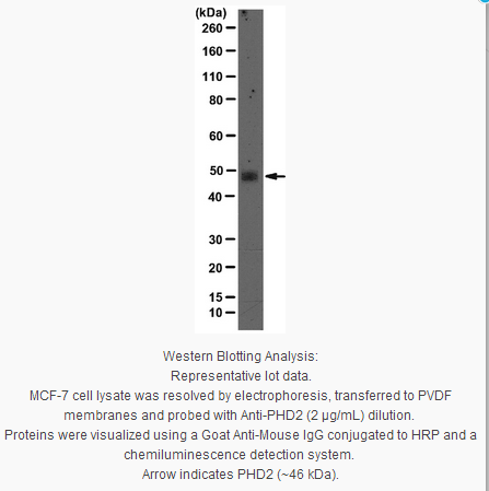
Evaluated by Western Blot in MCF-7 cell lysate.
Western Blot Analysis: 2 µg/ml of this antibody detected PHD2 on 10 µg of MCF-7 cell lysate.
Usage Statement
Unless otherwise stated in our catalog or other company documentation accompanying the product(s), our products are intended for research use only and are not to be used for any other purpose, which includes but is not limited to, unauthorized commercial uses, in vitro diagnostic uses, ex vivo or in vivo therapeutic uses or any type of consumption or application to humans or animals.
Storage and Shipping Information
Storage Conditions
Stable for 1 year at 2-8°C from date of receipt.
Packaging Information
Material Size
100 µg
原厂资料:
Key Spec Table
Species Reactivity
Key Applications
Host
Format
Antibody Type
H
WB
M
Purified
Monoclonal Antibody
Description
Catalogue Number
05-1327
Description
Anti-PHD2 Antibody, clone 76a
Alternate Names
EGL nine (C.elegans) homolog 1
egl nine-like protein 1
egl nine homolog 1 (C. elegans)
egl nine homolog 1
Prolyl hydroxylase domain-containing protein 2
Hypoxia-inducible factor prolyl hydroxylase 2
HIF-prolyl hydroxylase 2
HIF prolyl hydroxylase 2
Background Information
PHD2 (Prolyl hydroxylase domain-containing protein 2), also known as Hypoxia-inducible factor prolyl hydroxylase 2 (HIF-PH2 or HPH-2), is one of 4 PHD (PHD1-4) proteins which function as prolyl hydroxylases to effect oxygen homeostasis via hypoxia-inducible factor (HIF). This transcriptional complex is composed of an alpha-beta heterodimer that mediates a broad range of cellular and systemic responses to hypoxia. The PHD proteins hydroxylate HIF-1 alpha at 'Pro-402' and 'Pro-564', and HIF-2 alpha. The PHD2 isoform is the main down-regulator of HIFs for normal oxygen levels and mild hypoxia. When oxygen levels are normal, PHDs catalyze the hydroxylation of prolyl residues on HIF-1alpha. This post-translational modification targets this protein for proteasomal degradation via the von Hippel-Lindau ubiquitination complex.
Product Information
Format
Purified
Control
MCF-7 cell lysate
Presentation
Purified mouse monoclonal IgG1κ in buffer containing 0.1 M Tris-Glycine (pH 7.4, 150 mM NaCl) with 0.05% sodium azide.
Applications
Application
Use Anti-PHD2 Antibody, clone 76a (mouse monoclonal antibody) validated in WB to detect PHD2 also known as EGL nine (C.elegans) homolog 1, HIF prolyl hydroxylase 2, Hypoxia-inducible factor prolyl hydroxylase 2.
Key Applications
Western Blotting
Biological Information
Immunogen
Recombinant protein corresponding to human PHD2.
Epitope
Unknown
Clone
76a
Concentration
Please refer to the Certificate of Analysis for the lot-specific concentration.
FUNCTION: Catalyzes the post-translational formation of 4-hydroxyproline in hypoxia-inducible factor (HIF) alpha proteins. Hydroxylates HIF-1 alpha at 'Pro-402' and 'Pro-564', and HIF-2 alpha. Functions as a cellular oxygen sensor and, under normoxic conditions, targets HIF through the hydroxylation for proteasomal degradation via the von Hippel-Lindau ubiquitination complex.
ENZYME REGULATION: Following exposure to hypoxia, activated in HeLa cells but not in cardiovascular cells. Seems to be inhibited by ING4.
SUBUNIT STRUCTURE: Monomer. Interacts with ING4.
TISSUE SPECIFICITY: According to Ref.1 widely expressed with highest levels in skeletal muscle and heart, moderate levels in pancreas, brain (dopaminergic neurons of adult and fetal substantia nigra) and kidney, and lower levels in lung and liver. According to Ref.8 widely expressed with highest levels in brain, kidney and adrenal gland. Expressed in cardiac myocytes, aortic endothelial cells and coronary artery smooth muscle.
INVOLVEMENT IN DISEASE: Defects in EGLN1 are the cause of erythrocytosis familial type 3 (ECYT3) [MIM:609820]. ECYT3 is an autosomal dominant disorder characterized by increased serum red blood cell mass, elevated serum hemoglobin and hematocrit, and normal serum erythropoietin levels.
CAUTION: It was previously reported that this protein was the ortholog of rat SM-20. However, EGLN3 is now considered the true ortholog of rat SM-20 since it shows substantially greater similarity.
SEQUENCE CAUTION: The sequence AAK07536.1 differs from that shown. Reason: Frameshift at position 239.
Product Usage Statements
Quality Assurance
Evaluated by Western Blot in MCF-7 cell lysate.
Western Blot Analysis: 2 µg/ml of this antibody detected PHD2 on 10 µg of MCF-7 cell lysate.
Usage Statement
Unless otherwise stated in our catalog or other company documentation accompanying the product(s), our products are intended for research use only and are not to be used for any other purpose, which includes but is not limited to, unauthorized commercial uses, in vitro diagnostic uses, ex vivo or in vivo therapeutic uses or any type of consumption or application to humans or animals.


 京公网安备11010802025653 版权所有:北京逸优科技有限公司
