FAK is a 125 Kd protein that is recruited as a participant in focal adhesion dynamics between cells and has a role in motility and cell survival. FAK is a highly conserved, non-receptor tyrosine kinase originally identified as a substrate for the oncogene protein tyrosine kinase v-src (Guan, J L and Shalloway, D). This cytosolic kinase has been implicated in diverse cellular roles including cell locomotion, mitogen response and cell survival. FAK is typically located at structures known as focal adhesions, these are multi-protein structures that link the extracellular matrix (ECM) to the cytoplasmic cytoskeleton. Additional components of focal adhesions include actin, filamin, vinculin, talin, paxillin and tensin (Burridge, K, et al).
Product Information
Format
Purified
Control
HEK293 cell lysate
Presentation
Purified mouse monoclonal IgG1 in buffer containing 0.05% sodium azide.
Applications
Application
This Anti-Focal Adhesion Kinase Antibody, clone BLAb2H7 is validated for use in WB, IP for the detection of Focal Adhesion Kinase.
Key Applications
Western Blotting
Immunoprecipitation
Application Notes
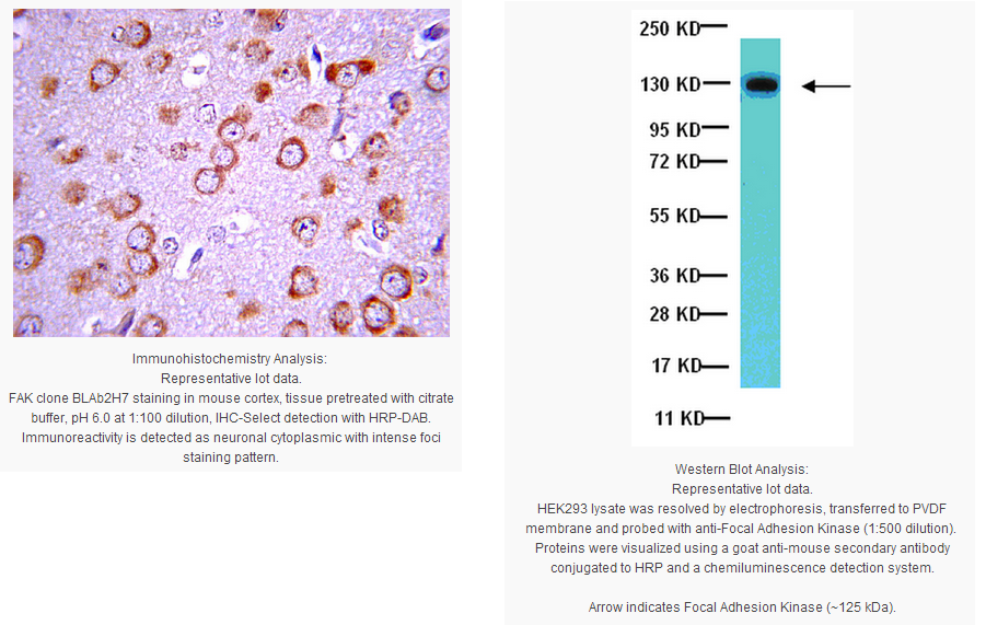
Immunohistochemistry Analysis: A previous lot detected Focal Adhesion Kinase at 1:100 dilution in mouse cortex tissue.
Biological Information
Immunogen
Recombinant fragment derived from the amino terminal region of chicken FAK, amino acid residues 103 to 553.
Epitope
Terminal
Clone
BLAb2H7
Concentration
Please refer to the Certificate of Analysis for the lot-specific concentration.
Host
Mouse
Specificity
This antibody recognizes Focal Adhesion Kinase
Isotype
IgG1
Species Reactivity
Human Chicken Rat Mouse
Species Reactivity Note
Demonstrated to react with human, mouse, rat and chicken.
FUNCTION:Non-receptor protein-tyrosine kinase implicated in signaling pathways involved in cell motility, proliferation and apoptosis. Activated by tyrosine-phosphorylation in response to either integrin clustering induced by cell adhesion or antibody cross-linking, or via G-protein coupled receptor (GPCR) occupancy by ligands such as bombesin or lysophosphatidic acid, or via LDL receptor occupancy. Plays a potential role in oncogenic transformations resulting in increased kinase activity. CATALYTIC ACTIVITY:ATP + a [protein]-L-tyrosine = ADP + a [protein]-L-tyrosine phosphate. SUBUNIT STRUCTURE:Interacts with CAS family members and with GIT1, SORBS1 and BCAR3. Interacts with RGNEF and SHB By similarity. Interacts with TGFB1I1 and STEAP4. SUBCELLULAR LOCATION:Cell junction › focal adhesion. Cell membrane; Peripheral membrane protein; Cytoplasmic side. Note: Constituent of focal adhesions. TISSUE SPECIFICITY:Expressed in all organs tested, in lymphoid cell lines, but most abundantly in brain. DOMAIN:The first Pro-rich domain interacts with the SH3 domain of CRK-associated substrate (BCAR1) and CASL By similarity.
The carboxy-terminal region is the site of focal adhesion targeting (FAT) sequence which mediates the localization of FAK1 to focal adhesions. PTM:Phosphorylated on 6 tyrosine residues upon activation. SEQUENCE SIMILARITIES:Belongs to the protein kinase superfamily. Tyr protein kinase family. FAK subfamily.
Contains 1 FERM domain.
Contains 1 protein kinase domain.
Product Usage Statements
Quality Assurance
Evaluated by Western Blotting in HEK293 cell lysate.
Western Blotting Analysis: 0.2 µg/mL of this antibody detected Focal Adhesion Kinase in 10 µg of HEK293 cell lysate.
Usage Statement
Unless otherwise stated in our catalog or other company documentation accompanying the product(s), our products are intended for research use only and are not to be used for any other purpose, which includes but is not limited to, unauthorized commercial uses, in vitro diagnostic uses, ex vivo or in vivo therapeutic uses or any type of consumption or application to humans or animals.
Storage and Shipping Information
Storage Conditions
Stable for 1 year at -20°C from date of receipt. Handling Recommendations: Upon first thaw, and prior to removing the cap, centrifuge the vial and gently mix the solution. Aliquot into microcentrifuge tubes and store at -20°C. Avoid repeated freeze/thaw cycles, which may damage IgG and affect product performance.
FAK is a 125 Kd protein that is recruited as a participant in focal adhesion dynamics between cells and has a role in motility and cell survival. FAK is a highly conserved, non-receptor tyrosine kinase originally identified as a substrate for the oncogene protein tyrosine kinase v-src (Guan, J L and Shalloway, D). This cytosolic kinase has been implicated in diverse cellular roles including cell locomotion, mitogen response and cell survival. FAK is typically located at structures known as focal adhesions, these are multi-protein structures that link the extracellular matrix (ECM) to the cytoplasmic cytoskeleton. Additional components of focal adhesions include actin, filamin, vinculin, talin, paxillin and tensin (Burridge, K, et al).
Product Information
Format
Purified
Control
HEK293 cell lysate
Presentation
Purified mouse monoclonal IgG1 in buffer containing 0.05% sodium azide.
Applications
Application
This Anti-Focal Adhesion Kinase Antibody, clone BLAb2H7 is validated for use in WB, IP for the detection of Focal Adhesion Kinase.
Key Applications
Western Blotting
Immunoprecipitation
Application Notes
Immunohistochemistry Analysis: A previous lot detected Focal Adhesion Kinase at 1:100 dilution in mouse cortex tissue.
Biological Information
Immunogen
Recombinant fragment derived from the amino terminal region of chicken FAK, amino acid residues 103 to 553.
Epitope
Terminal
Clone
BLAb2H7
Concentration
Please refer to the Certificate of Analysis for the lot-specific concentration.
Host
Mouse
Specificity
This antibody recognizes Focal Adhesion Kinase
Isotype
IgG1
Species Reactivity
Human Chicken Rat Mouse
Species Reactivity Note
Demonstrated to react with human, mouse, rat and chicken.
FUNCTION:Non-receptor protein-tyrosine kinase implicated in signaling pathways involved in cell motility, proliferation and apoptosis. Activated by tyrosine-phosphorylation in response to either integrin clustering induced by cell adhesion or antibody cross-linking, or via G-protein coupled receptor (GPCR) occupancy by ligands such as bombesin or lysophosphatidic acid, or via LDL receptor occupancy. Plays a potential role in oncogenic transformations resulting in increased kinase activity. CATALYTIC ACTIVITY:ATP + a [protein]-L-tyrosine = ADP + a [protein]-L-tyrosine phosphate. SUBUNIT STRUCTURE:Interacts with CAS family members and with GIT1, SORBS1 and BCAR3. Interacts with RGNEF and SHB By similarity. Interacts with TGFB1I1 and STEAP4. SUBCELLULAR LOCATION:Cell junction › focal adhesion. Cell membrane; Peripheral membrane protein; Cytoplasmic side. Note: Constituent of focal adhesions. TISSUE SPECIFICITY:Expressed in all organs tested, in lymphoid cell lines, but most abundantly in brain. DOMAIN:The first Pro-rich domain interacts with the SH3 domain of CRK-associated substrate (BCAR1) and CASL By similarity.
The carboxy-terminal region is the site of focal adhesion targeting (FAT) sequence which mediates the localization of FAK1 to focal adhesions. PTM:Phosphorylated on 6 tyrosine residues upon activation. SEQUENCE SIMILARITIES:Belongs to the protein kinase superfamily. Tyr protein kinase family. FAK subfamily.
Contains 1 FERM domain.
Contains 1 protein kinase domain.
Product Usage Statements
Quality Assurance
Evaluated by Western Blotting in HEK293 cell lysate.
Western Blotting Analysis: 0.2 µg/mL of this antibody detected Focal Adhesion Kinase in 10 µg of HEK293 cell lysate.
Usage Statement
Unless otherwise stated in our catalog or other company documentation accompanying the product(s), our products are intended for research use only and are not to be used for any other purpose, which includes but is not limited to, unauthorized commercial uses, in vitro diagnostic uses, ex vivo or in vivo therapeutic uses or any type of consumption or application to humans or animals.
Storage and Shipping Information
Storage Conditions
Stable for 1 year at -20°C from date of receipt. Handling Recommendations: Upon first thaw, and prior to removing the cap, centrifuge the vial and gently mix the solution. Aliquot into microcentrifuge tubes and store at -20°C. Avoid repeated freeze/thaw cycles, which may damage IgG and affect product performance.


 京公网安备11010802025653 版权所有:北京逸优科技有限公司
