v-ErbB was identified as the viral counterpart of the EGF Receptor, and its relatives have adopted this nomenclature (or the HER nomenclature for human proteins). These proteins are activated by Heregulin, Neuregulin and Amphiregulin. ErbB-2/HER-2, also known as neu, is amplified in large numbers of highly aggressive breast cancers, but all the family members are capable of heterodimerization, and together activate downstream signaling pathways. ErbB-3 has a natural "substitution" at the codon which is an invariant Lys residue at the ATP-binding site of all protein kinases. It is therefore inactive as a kinase, and can only become phosphorylated by heterodimerization with another erbB-family member. All members of this family, when Tyr-phosphorylated, can nucleate the formation of signaling complexes through adapter proteins.
Product Information
Format
Affinity Purified
Control
A431 Cell Lysate
Presentation
Purified mouse monoclonal IgG1 is presented in phosphate buffered saline containing 10mM sodium azide and 1mg/ml bovine serum albumin. We recommend that each laboratory determine an optimum working titre for use in its particular application.
Applications
Application
Anti-erbB-2 Antibody, clone N3/D10 (Cytoplasmic Domain) detects level of erbB-2 & has been published & validated for use in WB, IC, IH(P), FC.
Key Applications
Western Blotting
Flow Cytometry
Immunohistochemistry (Paraffin)
Immunocytochemistry
Application Notes
Immunocytochemistry: A previous lot of this antibody was used in immunocytochemistry on frozen sections.
Flow Cytometry: A previous lot of this antibody was used with FC.
Optimal working dilutions must be determined by the end user.
Immunohistochemistry(paraffin): Representative testing from a previous lot.
Optimal Staining of erbB-2/HER-2 Monoclonal Antibody: Breast Cancer
Biological Information
Immunogen
Synthetic peptide:
Cys-Lys-Gly-Thr-Pro-Thr-Ala-Glu-Asp-Pro-Glu-Tyr-Leu-Gly-Leu-Asp-Val-Pro-Val (amino acids 1238-1255) selected from the cytoplasmic domain of human c-erbB-2 protein.
Epitope
a.a. 1238-1255, Cytoplasmic Domain
Clone
N3/D10
Concentration
Please refer to the Certificate of Analysis for the lot-specific concentration.
Host
Mouse
Specificity
The antibody reacts with c-erbB-2 which is a membrane associated oncoprotein that has considerable sequence homology with EGFR. Membrane preparations were made from SK-BR-3 mammary adenocarcinoma and A431 epidermoid carcinoma cell lines. SK-BR-3 expresses high levels of c-erbB-2 whilst A431 expresses high levels of EGFR.
The antibody has been used to detect c-erbB-2 in paraffin-embedded material from breast tumours.
This gene encodes a member of the epidermal growth factor (EGF) receptor family of receptor tyrosine kinases. This protein has no ligand binding domain of its own and therefore cannot bind growth factors. However, it does bind tightly to other ligand-bound EGF receptor family members to form a heterodimer, stabilizing ligand binding and enhancing kinase-mediated activation of downstream signalling pathways, such as those involving mitogen-activated protein kinase and phosphatidylinositol-3 kinase. Allelic variations at amino acid positions 654 and 655 of isoform a (positions 624 and 625 of isoform b) have been reported, with the most common allele, Ile654/Ile655, shown here. Amplification and/or overexpression of this gene has been reported in numerous cancers, Including breast and ovarian tumors. Alternative splicing results in several additional transcript variants, some encoding different isoforms and others that have not been fully characterized.[provided by RefSeq].
FUNCTION: Essential component of a neuregulin-receptor complex, although neuregulins do not interact with it alone. GP30 is a potential ligand for this receptor. Not activated by EGF, TGF-alpha and amphiregulin.
CATALYTIC ACTIVITY: ATP + a [protein]-L-tyrosine = ADP + a [protein]-L-tyrosine phosphate.
SUBUNIT: Heterodimer with each of the other ERBB receptors (Potential). Interacts with PRKCABP and PLXNB1. Part of a complex with EGFR and either PIK3C2A or PIK3C2B. May interact with PIK3C2B when phosphorylated on Tyr-1196. Interacts with MEMO when phosphorylated on Tyr-1248. Interacts with MUC1. Stimulation by heregulin (HRG) in breast cancer cell lines induces binding of MUC1 with gamma-catenin.
SUBCELLULAR LOCATION: Membrane; Single-pass type I membrane protein.
PTM: Ligand-binding increases phosphorylation on tyrosine residues (By similarity).
POLYMORPHISM: There are fours alleles due to the variations in positions 654 and 655. Allele B1 (Ile-654/Ile-655) has a frequency of 0.782; allele B2 (Ile-654/Val-655) has a frequency of 0.206; allele B3 (Val-654/Val-655) has a frequency of 0.012.
SIMILARITY: Belongs to the protein kinase superfamily. Tyr protein kinase family. EGF receptor subfamily.
SIMILARITY: Contains 1 protein kinase domain.
WEB RESOURCE: Name=Atlas of Genetics and Cytogenetics in Oncology and Haematology; URL="http://atlasgeneticsoncology.org/Genes/ERBB2ID162ch17q11.html";.
WEB RESOURCE: Name=Wikipedia; Note=ERBB2 entry; URL="http://en.wikipedia.org/wiki/ERBB2";.
Product Usage Statements
Quality Assurance
Routinely evaluated by Western Blot on A431 lysates.
Western Blot Analysis: 1:500 dilution of this lot detected C-ERBB-2 on 10 μg of A431 lysates.
Usage Statement
Unless otherwise stated in our catalog or other company documentation accompanying the product(s), our products are intended for research use only and are not to be used for any other purpose, which includes but is not limited to, unauthorized commercial uses, in vitro diagnostic uses, ex vivo or in vivo therapeutic uses or any type of consumption or application to humans or animals.
Storage and Shipping Information
Storage Conditions
For use within 1 month of purchase store at 2-8°C, for long term storage aliquot antibody into small volumes and store at -20°C for up to 1 year from date of receipt.
Handling Recommendations: Upon first thaw, and prior to removing the cap, centrifuge the vial and gently mix the solution. Aliquot into microcentrifuge tubes and store at -20°C. Avoid repeated freeze/thaw cycles, which may damage IgG and affect product performance.
v-ErbB was identified as the viral counterpart of the EGF Receptor, and its relatives have adopted this nomenclature (or the HER nomenclature for human proteins). These proteins are activated by Heregulin, Neuregulin and Amphiregulin. ErbB-2/HER-2, also known as neu, is amplified in large numbers of highly aggressive breast cancers, but all the family members are capable of heterodimerization, and together activate downstream signaling pathways. ErbB-3 has a natural "substitution" at the codon which is an invariant Lys residue at the ATP-binding site of all protein kinases. It is therefore inactive as a kinase, and can only become phosphorylated by heterodimerization with another erbB-family member. All members of this family, when Tyr-phosphorylated, can nucleate the formation of signaling complexes through adapter proteins.
Product Information
Format
Affinity Purified
Control
A431 Cell Lysate
Presentation
Purified mouse monoclonal IgG1 is presented in phosphate buffered saline containing 10mM sodium azide and 1mg/ml bovine serum albumin. We recommend that each laboratory determine an optimum working titre for use in its particular application.
Applications
Application
Anti-erbB-2 Antibody, clone N3/D10 (Cytoplasmic Domain) detects level of erbB-2 & has been published & validated for use in WB, IC, IH(P), FC.
Key Applications
Western Blotting
Flow Cytometry
Immunohistochemistry (Paraffin)
Immunocytochemistry
Application Notes
Immunocytochemistry: A previous lot of this antibody was used in immunocytochemistry on frozen sections.
Flow Cytometry: A previous lot of this antibody was used with FC.
Optimal working dilutions must be determined by the end user.
Immunohistochemistry(paraffin): Representative testing from a previous lot.
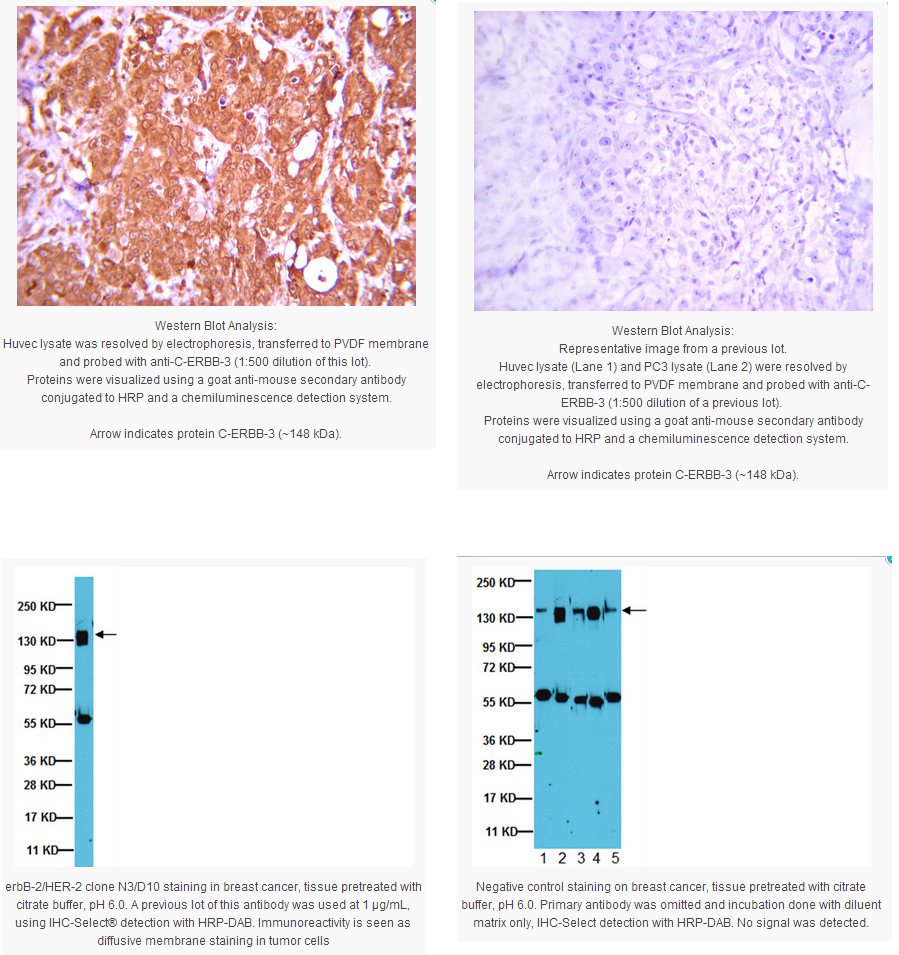
Optimal Staining of erbB-2/HER-2 Monoclonal Antibody: Breast Cancer
Biological Information
Immunogen
Synthetic peptide:
Cys-Lys-Gly-Thr-Pro-Thr-Ala-Glu-Asp-Pro-Glu-Tyr-Leu-Gly-Leu-Asp-Val-Pro-Val (amino acids 1238-1255) selected from the cytoplasmic domain of human c-erbB-2 protein.
Epitope
a.a. 1238-1255, Cytoplasmic Domain
Clone
N3/D10
Concentration
Please refer to the Certificate of Analysis for the lot-specific concentration.
Host
Mouse
Specificity
The antibody reacts with c-erbB-2 which is a membrane associated oncoprotein that has considerable sequence homology with EGFR. Membrane preparations were made from SK-BR-3 mammary adenocarcinoma and A431 epidermoid carcinoma cell lines. SK-BR-3 expresses high levels of c-erbB-2 whilst A431 expresses high levels of EGFR.
The antibody has been used to detect c-erbB-2 in paraffin-embedded material from breast tumours.
This gene encodes a member of the epidermal growth factor (EGF) receptor family of receptor tyrosine kinases. This protein has no ligand binding domain of its own and therefore cannot bind growth factors. However, it does bind tightly to other ligand-bound EGF receptor family members to form a heterodimer, stabilizing ligand binding and enhancing kinase-mediated activation of downstream signalling pathways, such as those involving mitogen-activated protein kinase and phosphatidylinositol-3 kinase. Allelic variations at amino acid positions 654 and 655 of isoform a (positions 624 and 625 of isoform b) have been reported, with the most common allele, Ile654/Ile655, shown here. Amplification and/or overexpression of this gene has been reported in numerous cancers, Including breast and ovarian tumors. Alternative splicing results in several additional transcript variants, some encoding different isoforms and others that have not been fully characterized.[provided by RefSeq].
FUNCTION: Essential component of a neuregulin-receptor complex, although neuregulins do not interact with it alone. GP30 is a potential ligand for this receptor. Not activated by EGF, TGF-alpha and amphiregulin.
CATALYTIC ACTIVITY: ATP + a [protein]-L-tyrosine = ADP + a [protein]-L-tyrosine phosphate.
SUBUNIT: Heterodimer with each of the other ERBB receptors (Potential). Interacts with PRKCABP and PLXNB1. Part of a complex with EGFR and either PIK3C2A or PIK3C2B. May interact with PIK3C2B when phosphorylated on Tyr-1196. Interacts with MEMO when phosphorylated on Tyr-1248. Interacts with MUC1. Stimulation by heregulin (HRG) in breast cancer cell lines induces binding of MUC1 with gamma-catenin.
SUBCELLULAR LOCATION: Membrane; Single-pass type I membrane protein.
PTM: Ligand-binding increases phosphorylation on tyrosine residues (By similarity).
POLYMORPHISM: There are fours alleles due to the variations in positions 654 and 655. Allele B1 (Ile-654/Ile-655) has a frequency of 0.782; allele B2 (Ile-654/Val-655) has a frequency of 0.206; allele B3 (Val-654/Val-655) has a frequency of 0.012.
SIMILARITY: Belongs to the protein kinase superfamily. Tyr protein kinase family. EGF receptor subfamily.
SIMILARITY: Contains 1 protein kinase domain.
WEB RESOURCE: Name=Atlas of Genetics and Cytogenetics in Oncology and Haematology; URL="http://atlasgeneticsoncology.org/Genes/ERBB2ID162ch17q11.html";.
WEB RESOURCE: Name=Wikipedia; Note=ERBB2 entry; URL="http://en.wikipedia.org/wiki/ERBB2";.
Product Usage Statements
Quality Assurance
Routinely evaluated by Western Blot on A431 lysates.
Western Blot Analysis: 1:500 dilution of this lot detected C-ERBB-2 on 10 μg of A431 lysates.
Usage Statement
Unless otherwise stated in our catalog or other company documentation accompanying the product(s), our products are intended for research use only and are not to be used for any other purpose, which includes but is not limited to, unauthorized commercial uses, in vitro diagnostic uses, ex vivo or in vivo therapeutic uses or any type of consumption or application to humans or animals.
Storage and Shipping Information
Storage Conditions
For use within 1 month of purchase store at 2-8°C, for long term storage aliquot antibody into small volumes and store at -20°C for up to 1 year from date of receipt.
Handling Recommendations: Upon first thaw, and prior to removing the cap, centrifuge the vial and gently mix the solution. Aliquot into microcentrifuge tubes and store at -20°C. Avoid repeated freeze/thaw cycles, which may damage IgG and affect product performance.


 京公网安备11010802025653 版权所有:北京逸优科技有限公司
