IGF-I and -II signal through the IGF-I Receptor, which is homologous to the Insulin Receptor. The high-affinity IGF-II Receptor does not play a direct role in signaling, but regulates the concentration of free IGF-II. The IGFs are involved in skeletal growth, and are essential for prevention of apoptosis. Serum levels of free IGFs are kept low by the action of IGF binding proteins (IGFBPs), which sequester the IGFs. Overexpression of IGFBPs may induce apoptosis, presumably by reduction of free IGF; IGFBP levels are also altered in some cancers. The IGF-I Receptor is not as mitogenic as some other growth factor receptors, but its ability to activate the PI3 Kinase pathway, through the Insulin Receptor Substrate (IRS) proteins, is critical for mediating cell survival.
Product Information
Format
Affinity Purified
Control
C2C12 Cell Lysate
Presentation
Purified mouse monoclonal IgG2b in 10mM PBS, pH 7.4, with 0.2% BSA and 0.09% sodium azide.
Applications
Application
Anti-IGF-IR Antibody, β-subunit, clone 1-2 is an antibody against IGF-IR for use in WB, IH(P).
Key Applications
Western Blotting
Immunohistochemistry (Paraffin)
Application Notes
Immunohistochemistry(paraffin): Representative images from a previous lot.
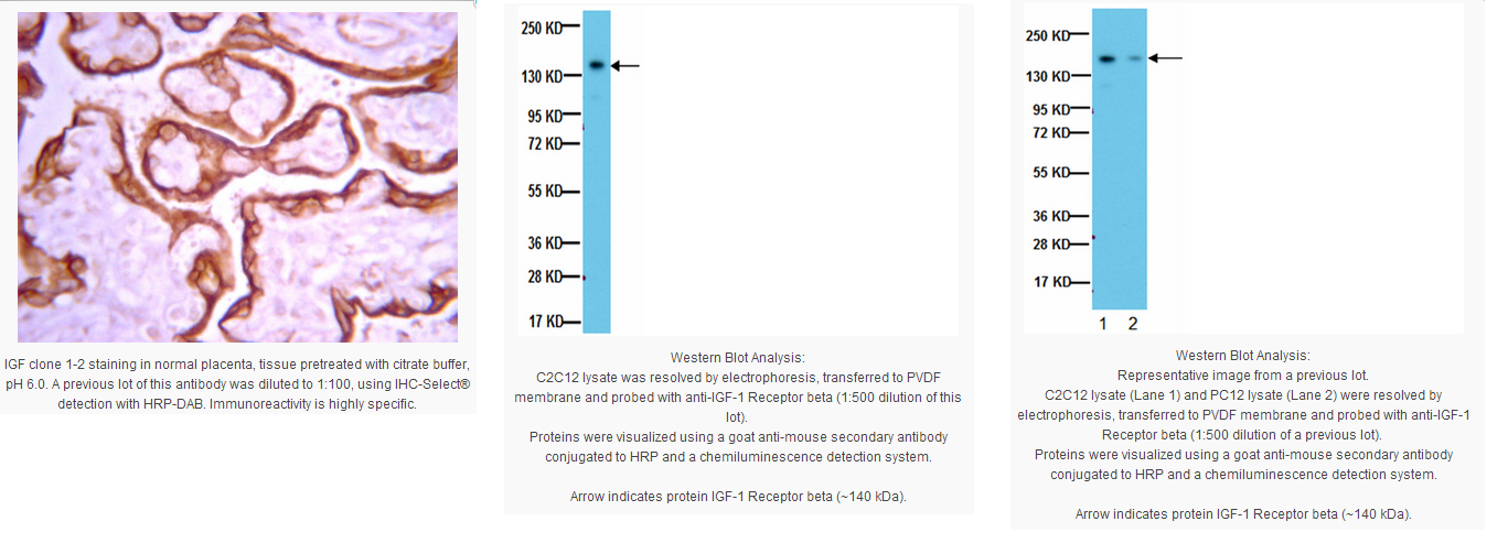
Optimal Staining of IGF Monoclonal Antibody: Placenta
Western Blot: A previous lot of this antibody was used at 1-2 µg/mL for 2 hours at room temperature.
Optimal working dilutions must be determined by end user.
Biological Information
Immunogen
A 15-mer peptide corresponding to a.a. 1323-1337 (Tyr-RKNERALPLPQSSTC) from the C-terminal of β-subunit of human IGF-1R.
Epitope
Between a.a. 1323-1337 of IGF-1R
Clone
1-2
Concentration
Please refer to the Certificate of Analysis for the lot-specific concentration.
Host
Mouse
Specificity
Recognizes the β-subunit of type I Insulin-like Growth Factor Receptor (IGF-1R). It shows no cross-reaction with insulin receptor. Its epitope localizes between a.a. 1323-1337 of IGF-1R. Both IGF-1R and insulin receptor are synthesized as a single polypeptide which is glycosylated and proteolytically cleaved to give the α- and β-subunits, which are disulfide-linked in a β-α-α-β configuration in the mature receptor. The α-subunit is completely extracellular, while the β-subunit spans membrane.
Isotype
IgG2b
Species Reactivity
Human Mouse Rat
Species Reactivity Note
Mouse and rat. Expected to react with human based on sequence homology.
This receptor binds insulin-like growth factor with a high affinity. It has tyrosine kinase activity. The insulin-like growth factor I receptor plays a critical role in transformation events. Cleavage of the precursor generates alpha and beta subunits. It is highly overexpressed in most malignant tissues where it functions as an anti-apoptotic agent by enhancing cell survival. [provided by RefSeq]
FUNCTION: This receptor binds insulin-like growth factor 1 (IGF1) with a high affinity and IGF2 with a lower affinity. It has a tyrosine-protein kinase activity, which is necessary for the activation of the IGF1-stimulated downstream signaling cascade.
CATALYTIC ACTIVITY: ATP + a [protein]-L-tyrosine = ADP + a [protein]-L-tyrosine phosphate.
ENZYME REGULATION: Autophosphorylation activates the kinase activity.
SUBUNIT: Tetramer of 2 alpha and 2 beta chains linked by disulfide bonds. The alpha chains contribute to the formation of the ligand-binding domain, while the beta chain carries the kinase domain. Interacts with PIK3R1 and with the PTB/PID domains of IRS1 and SHC1 in vitro when autophosphorylated on tyrosine residues.
SUBCELLULAR LOCATION: Membrane; Single-pass type I membrane protein.
TISSUE SPECIFICITY: Expressed in a variety of tissues.
PTM: The cytoplasmic domain of the beta subunit is autophosphorylated on tyrosine residues in response to insulin-like growth factor I (IGF I).
PTM: Phosphorylation of Tyr-980 is required for IRS1- and SHC1-binding.
DISEASE: Defects in IGF1R may be a cause in some cases of resistance to insulin-like growth factor 1 (IGF1 resistance) [MIM:270450]. IGF1 resistance is a gowth deficiency disorder characterized by intrauterine growth retardation and poor postnatal growth accompanied with increased plasma IGF1.
SIMILARITY: Belongs to the protein kinase superfamily. Tyr protein kinase family. Insulin receptor subfamily.
SIMILARITY: Contains 3 fibronectin type-III domains.
SIMILARITY: Contains 1 protein kinase domain.
Product Usage Statements
Quality Assurance
Routinely evaluated by Western Blot on C2C12 lysates.
Western Blot Analysis: 1:500 dilution of this lot detected IGF-1 Receptor beta on 10 μg of C2C12 lysates.
Usage Statement
Unless otherwise stated in our catalog or other company documentation accompanying the product(s), our products are intended for research use only and are not to be used for any other purpose, which includes but is not limited to, unauthorized commercial uses, in vitro diagnostic uses, ex vivo or in vivo therapeutic uses or any type of consumption or application to humans or animals.
Storage and Shipping Information
Storage Conditions
Stable at 2-8°C in undiluted aliquots for up to 1 year from date of receipt.
Packaging Information
Material Size
100 µg
原厂资料:
Key Spec Table
Species Reactivity
Key Applications
Host
Format
Antibody Type
H, M, R
WB, IH(P)
M
Affinity Purified
Monoclonal Antibody
Description
Catalogue Number
05-1106
Replaces
MAB1123
Description
Anti-IGF-IR Antibody, β-subunit, clone 1-2
Background Information
IGF-I and -II signal through the IGF-I Receptor, which is homologous to the Insulin Receptor. The high-affinity IGF-II Receptor does not play a direct role in signaling, but regulates the concentration of free IGF-II. The IGFs are involved in skeletal growth, and are essential for prevention of apoptosis. Serum levels of free IGFs are kept low by the action of IGF binding proteins (IGFBPs), which sequester the IGFs. Overexpression of IGFBPs may induce apoptosis, presumably by reduction of free IGF; IGFBP levels are also altered in some cancers. The IGF-I Receptor is not as mitogenic as some other growth factor receptors, but its ability to activate the PI3 Kinase pathway, through the Insulin Receptor Substrate (IRS) proteins, is critical for mediating cell survival.
Product Information
Format
Affinity Purified
Control
C2C12 Cell Lysate
Presentation
Purified mouse monoclonal IgG2b in 10mM PBS, pH 7.4, with 0.2% BSA and 0.09% sodium azide.
Applications
Application
Anti-IGF-IR Antibody, β-subunit, clone 1-2 is an antibody against IGF-IR for use in WB, IH(P).
Key Applications
Western Blotting
Immunohistochemistry (Paraffin)
Application Notes
Immunohistochemistry(paraffin): Representative images from a previous lot.
Optimal Staining of IGF Monoclonal Antibody: Placenta
Western Blot: A previous lot of this antibody was used at 1-2 µg/mL for 2 hours at room temperature.
Optimal working dilutions must be determined by end user.
Biological Information
Immunogen
A 15-mer peptide corresponding to a.a. 1323-1337 (Tyr-RKNERALPLPQSSTC) from the C-terminal of β-subunit of human IGF-1R.
Epitope
Between a.a. 1323-1337 of IGF-1R
Clone
1-2
Concentration
Please refer to the Certificate of Analysis for the lot-specific concentration.
Host
Mouse
Specificity
Recognizes the β-subunit of type I Insulin-like Growth Factor Receptor (IGF-1R). It shows no cross-reaction with insulin receptor. Its epitope localizes between a.a. 1323-1337 of IGF-1R. Both IGF-1R and insulin receptor are synthesized as a single polypeptide which is glycosylated and proteolytically cleaved to give the α- and β-subunits, which are disulfide-linked in a β-α-α-β configuration in the mature receptor. The α-subunit is completely extracellular, while the β-subunit spans membrane.
Isotype
IgG2b
Species Reactivity
Human Mouse Rat
Species Reactivity Note
Mouse and rat. Expected to react with human based on sequence homology.
This receptor binds insulin-like growth factor with a high affinity. It has tyrosine kinase activity. The insulin-like growth factor I receptor plays a critical role in transformation events. Cleavage of the precursor generates alpha and beta subunits. It is highly overexpressed in most malignant tissues where it functions as an anti-apoptotic agent by enhancing cell survival. [provided by RefSeq]
FUNCTION: This receptor binds insulin-like growth factor 1 (IGF1) with a high affinity and IGF2 with a lower affinity. It has a tyrosine-protein kinase activity, which is necessary for the activation of the IGF1-stimulated downstream signaling cascade.
CATALYTIC ACTIVITY: ATP + a [protein]-L-tyrosine = ADP + a [protein]-L-tyrosine phosphate.
ENZYME REGULATION: Autophosphorylation activates the kinase activity.
SUBUNIT: Tetramer of 2 alpha and 2 beta chains linked by disulfide bonds. The alpha chains contribute to the formation of the ligand-binding domain, while the beta chain carries the kinase domain. Interacts with PIK3R1 and with the PTB/PID domains of IRS1 and SHC1 in vitro when autophosphorylated on tyrosine residues.
SUBCELLULAR LOCATION: Membrane; Single-pass type I membrane protein.
TISSUE SPECIFICITY: Expressed in a variety of tissues.
PTM: The cytoplasmic domain of the beta subunit is autophosphorylated on tyrosine residues in response to insulin-like growth factor I (IGF I).
PTM: Phosphorylation of Tyr-980 is required for IRS1- and SHC1-binding.
DISEASE: Defects in IGF1R may be a cause in some cases of resistance to insulin-like growth factor 1 (IGF1 resistance) [MIM:270450]. IGF1 resistance is a gowth deficiency disorder characterized by intrauterine growth retardation and poor postnatal growth accompanied with increased plasma IGF1.
SIMILARITY: Belongs to the protein kinase superfamily. Tyr protein kinase family. Insulin receptor subfamily.
SIMILARITY: Contains 3 fibronectin type-III domains.
SIMILARITY: Contains 1 protein kinase domain.
Product Usage Statements
Quality Assurance
Routinely evaluated by Western Blot on C2C12 lysates.
Western Blot Analysis: 1:500 dilution of this lot detected IGF-1 Receptor beta on 10 μg of C2C12 lysates.
Usage Statement
Unless otherwise stated in our catalog or other company documentation accompanying the product(s), our products are intended for research use only and are not to be used for any other purpose, which includes but is not limited to, unauthorized commercial uses, in vitro diagnostic uses, ex vivo or in vivo therapeutic uses or any type of consumption or application to humans or animals.
Storage and Shipping Information
Storage Conditions
Stable at 2-8°C in undiluted aliquots for up to 1 year from date of receipt.


 京公网安备11010802025653 版权所有:北京逸优科技有限公司
