The insulin receptor is a tyrosine kinase receptor that when bound to insulin, initiates multiple signal transduction pathways, including JNK, PI 3-kinase, Akt and PKC. Pharmacological intervention of these Insulin R-dependent pathways is of great interest for the treatment of insulin resistance, obesity and diabetes. The Insulin Receptor (IR) is synthesized as a single polypeptide, which is subsequently cleaved to generate an extracellular α-chain and a transmembrane and intracellular β-chain, tethered together by disulfide bonds. The β-chain has multiple tyrosine phosphorylation sites, including three autophosphorylation sites at its activation loop. The overall structure of the IR is highly homologous to the IGF-I Receptor, except in their c-termini, where the two proteins diverge somewhat. The IR signals primarily by phosphorylating the Insulin Receptor Substrate (IRS) family of proteins, which creates docking sites for SH2-domain containing proteins. Insulin signaling is highly dependent on the PI3 Kinase pathway and Akt, which appear to mediate the functions of insulin.
Product Information
Format
Purified
Control
NIH/3T3 cell lysate
Presentation
Purified mouse monoclonal IgG1 in buffer containing 10 mM PBS, pH 7.4, with 0.2% BSA and 0.09% sodium azide.
Applications
Application
Anti-Insulin Receptor (β-Subunit) Antibody, clone CT-3 detects level of Insulin Receptor (β-Subunit) & has been published & validated for use in ELISA, EA, WB, IH(P), IH.
Key Applications
ELISA
Immunohistochemistry
Immunohistochemistry (Paraffin)
Western Blotting
Enzyme Assay
Application Notes
Immunohistochemistry (frozen and formalin/paraffin):
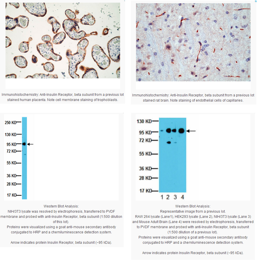
2-4ug/mL of a previous lot of this antibody was used in immunohistochemistry. Staining of formalin-fixed tissues requires boiling tissue sections in 10mM citrate buffer, pH 6.0, for 10-20 min followed by cooling at room temperature for 20 min.
Optimal working dilutions must be determined by end user.
ELISA: A previous lot of this antibody showed to be suitable as a capture antibody for ELISA.
Kinase Assay: A previous lot of this antibody was used for antibody-mediated capture on microplates.
Western Blot: A previous lot of this antibody was used at 1 ug/mL, for 2 hrs at room temperature.
Biological Information
Immunogen
Recombinant-fragment including the C-terminal 100 amino acids of human insulin receptor, β subunit.
Epitope
100 C-Terminal amino acids of human insulin receptor, β-subunit.
Clone
CT-3
Concentration
Please refer to the Certificate of Analysis for the lot-specific concentration.
Host
Mouse
Specificity
Recognizes a protein of 95 kDa, identified as the β-subunit of insulin receptor (IR). It is directed against the C-terminal region of β-subunit and shows no cross-reaction with IGF-receptors.
After removal of the precursor signal peptide, the insulin receptor precursor is post-translationally cleaved into two chains (alpha and beta) that are covalently linked. Binding of insulin to the insulin receptor (INSR) stimulates glucose uptake. Two transcript variants encoding different isoforms have been found for this gene. [provided by RefSeq]
FUNCTION: This receptor binds insulin and has a tyrosine-protein kinase activity. Isoform Short has a higher affinity for insulin. Mediates the metabolic functions of insulin. Binding to insulin stimulates association of the receptor with downstream mediators including IRS1 and phosphatidylinositol 3'-kinase (PI3K). Can activate PI3K either directly by binding to the p85 regulatory subunit, or indirectly via IRS1.
CATALYTIC ACTIVITY: ATP + a [protein]-L-tyrosine = ADP + a [protein]-L-tyrosine phosphate.
ENZYME REGULATION: Autophosphorylation activates the kinase activity.
SUBUNIT: Tetramer of 2 alpha and 2 beta chains linked by disulfide bonds. The alpha chains contribute to the formation of the ligand-binding domain, while the beta chains carry the kinase domain. Interacts with SORBS1 but dissociates from it following insulin stimulation. Binds SH2B2. Interacts with the PTB/PID domains of IRS1 and SHC1 in vitro when autophosphorylated on tyrosine residues. The sequences surrounding the phosphorylated NPXY motif contribute differentially to either IRS1 or SHC1 recognition. Interacts with the SH2 domains of the 85 kDa regulatory subunit of PI3K (PIK3R1) in vitro, when autophosphorylated on tyrosine residues. Interacts with SOCS7.
SUBCELLULAR LOCATION: Membrane; Single-pass type I membrane protein.
ALTERNATIVE PRODUCTS: 2 named isoforms [FASTA] produced by alternative
Product Usage Statements
Quality Assurance
Routinely evaluated by Western Blot on NIH/3T3 lysates.
Western Blot Analysis: 1:500 dilution of this lot detected Insulin Receptor, beta subunit on 10 μg of NIH/3T3 lysates.
Usage Statement
Unless otherwise stated in our catalog or other company documentation accompanying the product(s), our products are intended for research use only and are not to be used for any other purpose, which includes but is not limited to, unauthorized commercial uses, in vitro diagnostic uses, ex vivo or in vivo therapeutic uses or any type of consumption or application to humans or animals.
Storage and Shipping Information
Storage Conditions
Stable for 1 year at 2-8°C in undiluted aliquots from date of receipt.
The insulin receptor is a tyrosine kinase receptor that when bound to insulin, initiates multiple signal transduction pathways, including JNK, PI 3-kinase, Akt and PKC. Pharmacological intervention of these Insulin R-dependent pathways is of great interest for the treatment of insulin resistance, obesity and diabetes. The Insulin Receptor (IR) is synthesized as a single polypeptide, which is subsequently cleaved to generate an extracellular α-chain and a transmembrane and intracellular β-chain, tethered together by disulfide bonds. The β-chain has multiple tyrosine phosphorylation sites, including three autophosphorylation sites at its activation loop. The overall structure of the IR is highly homologous to the IGF-I Receptor, except in their c-termini, where the two proteins diverge somewhat. The IR signals primarily by phosphorylating the Insulin Receptor Substrate (IRS) family of proteins, which creates docking sites for SH2-domain containing proteins. Insulin signaling is highly dependent on the PI3 Kinase pathway and Akt, which appear to mediate the functions of insulin.
Product Information
Format
Purified
Control
NIH/3T3 cell lysate
Presentation
Purified mouse monoclonal IgG1 in buffer containing 10 mM PBS, pH 7.4, with 0.2% BSA and 0.09% sodium azide.
Applications
Application
Anti-Insulin Receptor (β-Subunit) Antibody, clone CT-3 detects level of Insulin Receptor (β-Subunit) & has been published & validated for use in ELISA, EA, WB, IH(P), IH.
Key Applications
ELISA
Immunohistochemistry
Immunohistochemistry (Paraffin)
Western Blotting
Enzyme Assay
Application Notes
Immunohistochemistry (frozen and formalin/paraffin):
2-4ug/mL of a previous lot of this antibody was used in immunohistochemistry. Staining of formalin-fixed tissues requires boiling tissue sections in 10mM citrate buffer, pH 6.0, for 10-20 min followed by cooling at room temperature for 20 min.
Optimal working dilutions must be determined by end user.
ELISA: A previous lot of this antibody showed to be suitable as a capture antibody for ELISA.
Kinase Assay: A previous lot of this antibody was used for antibody-mediated capture on microplates.
Western Blot: A previous lot of this antibody was used at 1 ug/mL, for 2 hrs at room temperature.
Biological Information
Immunogen
Recombinant-fragment including the C-terminal 100 amino acids of human insulin receptor, β subunit.
Epitope
100 C-Terminal amino acids of human insulin receptor, β-subunit.
Clone
CT-3
Concentration
Please refer to the Certificate of Analysis for the lot-specific concentration.
Host
Mouse
Specificity
Recognizes a protein of 95 kDa, identified as the β-subunit of insulin receptor (IR). It is directed against the C-terminal region of β-subunit and shows no cross-reaction with IGF-receptors.
After removal of the precursor signal peptide, the insulin receptor precursor is post-translationally cleaved into two chains (alpha and beta) that are covalently linked. Binding of insulin to the insulin receptor (INSR) stimulates glucose uptake. Two transcript variants encoding different isoforms have been found for this gene. [provided by RefSeq]
FUNCTION: This receptor binds insulin and has a tyrosine-protein kinase activity. Isoform Short has a higher affinity for insulin. Mediates the metabolic functions of insulin. Binding to insulin stimulates association of the receptor with downstream mediators including IRS1 and phosphatidylinositol 3'-kinase (PI3K). Can activate PI3K either directly by binding to the p85 regulatory subunit, or indirectly via IRS1.
CATALYTIC ACTIVITY: ATP + a [protein]-L-tyrosine = ADP + a [protein]-L-tyrosine phosphate.
ENZYME REGULATION: Autophosphorylation activates the kinase activity.
SUBUNIT: Tetramer of 2 alpha and 2 beta chains linked by disulfide bonds. The alpha chains contribute to the formation of the ligand-binding domain, while the beta chains carry the kinase domain. Interacts with SORBS1 but dissociates from it following insulin stimulation. Binds SH2B2. Interacts with the PTB/PID domains of IRS1 and SHC1 in vitro when autophosphorylated on tyrosine residues. The sequences surrounding the phosphorylated NPXY motif contribute differentially to either IRS1 or SHC1 recognition. Interacts with the SH2 domains of the 85 kDa regulatory subunit of PI3K (PIK3R1) in vitro, when autophosphorylated on tyrosine residues. Interacts with SOCS7.
SUBCELLULAR LOCATION: Membrane; Single-pass type I membrane protein.
ALTERNATIVE PRODUCTS: 2 named isoforms [FASTA] produced by alternative
Product Usage Statements
Quality Assurance
Routinely evaluated by Western Blot on NIH/3T3 lysates.
Western Blot Analysis: 1:500 dilution of this lot detected Insulin Receptor, beta subunit on 10 μg of NIH/3T3 lysates.
Usage Statement
Unless otherwise stated in our catalog or other company documentation accompanying the product(s), our products are intended for research use only and are not to be used for any other purpose, which includes but is not limited to, unauthorized commercial uses, in vitro diagnostic uses, ex vivo or in vivo therapeutic uses or any type of consumption or application to humans or animals.
Storage and Shipping Information
Storage Conditions
Stable for 1 year at 2-8°C in undiluted aliquots from date of receipt.


 京公网安备11010802025653 版权所有:北京逸优科技有限公司
