Protein phosphatase 1, regulatory (inhibitor) subunit 12A
Myosin phosphatase-targeting subunit 1
Protein phosphatase myosin-binding subunit
myosin phosphatase, target subunit 1
protein phosphatase 1 regulatory subunit 12A
target subunit 1
protein phosphatase 1 regulatory subunit 12A
Background Information
The major protein phosphatase-1 (PP1) is composed of 3 subunits with apparent molecular mass of 110 -130, 37 and 20 kDa. The 110-130 kDa component acts as a regulatory subunit known as myosin binding (MBS) or myosin phosphatase targeting subunit (MYPT). The N-terminal portion of MYPT binds to both PP1c-delta and phosphorylated myosin, thereby increasing myosin phosphatase activity. Two isoforms of MYPT have been identified (MYPT1 & MYPT2) and share 61% sequence identity. While MYPT1 is widely distributed in human tissues, MYPT2 is mostly detected in brain and heart.
Product Information
Format
Unpurified
Control
293T cell lysate
Presentation
Rabbit Monoclonal in buffer containing glycerol, BSA, and sodium azide.
Applications
Application
Please note that this product will not be available for sale after March 15, 2015. Please select one of the other antibodies against this target.
Key Applications
Flow Cytometry
Western Blotting
Immunocytochemistry
Immunoprecipitation
Application Notes
Immunocytochemistry Analysis: A 1:100 dilution from a representative lot was used in IC.
Immunoprecipitation Analysis: A 1:10-100 dilution from a representative lot was used in IP.
Flow Cytometry Analysis: A 1:10-100 dilution from a representative lot was used in FC.
Biological Information
Immunogen
Synthetic peptide corresponding to the C-terminus of human MYPT1/MYPT2.
Epitope
C-terminus
Clone
YE336
Host
Rabbit
Specificity
This antibody recognizes the C-terminus of MYPT1/MYPT2.
FUNCTION:Key regulator of protein phosphatase 1C (PPP1C). Mediates binding to myosin. As part of the PPP1C complex, involved in dephosphorylation of PLK1. SUBUNIT STRUCTURE:PP1 comprises a catalytic subunit, PPP1CA, PPP1CB or PPP1CC, and one or several targeting or regulatory subunits. PPP1R12A mediates binding to myosin. Interacts with ARHA and CIT By similarity. Binds PPP1R12B, ROCK1 and IL16. Interacts directly with PRKG1. Non-covalent dimer of 2 dimers; PRKG1-PRKG1 and PPP1R12A-PPP1R12A. Interacts with SMTNL1 By similarity. Interacts with PPP1CB; the interaction is direct. Interacts (when phosphorylated at Ser-445, Ser-472 and Ser-910) with 14-3-3. Interacts with ROCK1 and ROCK2. Interacts with isoform 1 and isoform 2 of ZIPK/DAPK3. Interacts with RAF1. SUBCELLULAR LOCATION:Cytoplasm. Note: Along actomyosin filaments and stress fibers.TISSUE SPECIFICITY:Expressed in striated muscles, specifically in type 2a fibers (at protein level). DEVELOPMENTAL STAGE:Induced by 2-fold during pregnancy, including in abdominus rectus muscle.DOMAIN:Heterotetramerization is mediated by the interaction between a coiled-coil of PRKG1 and the leucine/isoleucine zipper of PPP1R12A/MBS, the myosin-binding subunit of the myosin phosphatase By similarity.
The KVKF motif mediates interaction with PPP1CB. PTM:Phosphorylated by CIT (Rho-associated kinase) By similarity. Phosphorylated cooperatively by ROCK1 and CDC42BP on Thr-696. Phosphorylated on upon DNA damage, probably by ATM or ATR. In vitro, phosphorylation of Ser-695 by PKA and PKG appears to prevent phosphorylation of the inhibitory site Thr-696, probably mediated by PRKG1. Phosphorylation at Ser-445, Ser-472 and Ser-910 by NUAK1 promotes interaction with 14-3-3, leading to inhibit interaction with myosin light chain MLC2, preventing dephosphorylation of MLC2. May be phosphorylated at Thr-696 by DMPK; may inhibit the myosin phosphatase activity. Phosphorylated at Ser-473 by CDK1 during mitosis, creating docking sites for the POLO box domains of PLK1. Subsequently, PLK1 binds and phosphorylates PPP1R12A. SEQUENCE SIMILARITIES:Contains 6 ANK repeats. SEQUENCE CAUTION:The sequence AAH47898.1 differs from that shown. Reason: Contaminating sequence. Potential poly-A sequence.
The sequence AAH92481.1 differs from that shown. Reason: Contaminating sequence. Potential poly-A sequence.
Product Usage Statements
Quality Assurance
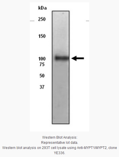
Evaluated by Western Blot in 293T cell lysate.
Western Blot Analysis: 1:5,000 dilution
of this antibody detected MYPT1/MYPT2 in 293T cell lysate.
Usage Statement
Unless otherwise stated in our catalog or other company documentation accompanying the product(s), our products are intended for research use only and are not to be used for any other purpose, which includes but is not limited to, unauthorized commercial uses, in vitro diagnostic uses, ex vivo or in vivo therapeutic uses or any type of consumption or application to humans or animals.
Storage and Shipping Information
Storage Conditions
Stable for 1 year at -20ºC from date of receipt.
Handling Recommendations: Upon first thaw, and prior to removing cap, centrifuge the vial and gently mix the solution. Aliquot into microcentrifuge tubes and store at -20ºC. Avoid freeze/thaw cycles, which may damage IgG and affect product performance.
Note: Variability in freezer temperatures below -20°C may cause glycerol containing solutions to become frozen during storage. Note: Variability in freezer temperatures below -20°C may cause glycerol containing solutions to become frozen during storage.
Protein phosphatase 1, regulatory (inhibitor) subunit 12A
Myosin phosphatase-targeting subunit 1
Protein phosphatase myosin-binding subunit
myosin phosphatase, target subunit 1
protein phosphatase 1 regulatory subunit 12A
target subunit 1
protein phosphatase 1 regulatory subunit 12A
Background Information
The major protein phosphatase-1 (PP1) is composed of 3 subunits with apparent molecular mass of 110 -130, 37 and 20 kDa. The 110-130 kDa component acts as a regulatory subunit known as myosin binding (MBS) or myosin phosphatase targeting subunit (MYPT). The N-terminal portion of MYPT binds to both PP1c-delta and phosphorylated myosin, thereby increasing myosin phosphatase activity. Two isoforms of MYPT have been identified (MYPT1 & MYPT2) and share 61% sequence identity. While MYPT1 is widely distributed in human tissues, MYPT2 is mostly detected in brain and heart.
Product Information
Format
Unpurified
Control
293T cell lysate
Presentation
Rabbit Monoclonal in buffer containing glycerol, BSA, and sodium azide.
Applications
Application
Please note that this product will not be available for sale after March 15, 2015. Please select one of the other antibodies against this target.
Key Applications
Flow Cytometry
Western Blotting
Immunocytochemistry
Immunoprecipitation
Application Notes
Immunocytochemistry Analysis: A 1:100 dilution from a representative lot was used in IC.
Immunoprecipitation Analysis: A 1:10-100 dilution from a representative lot was used in IP.
Flow Cytometry Analysis: A 1:10-100 dilution from a representative lot was used in FC.
Biological Information
Immunogen
Synthetic peptide corresponding to the C-terminus of human MYPT1/MYPT2.
Epitope
C-terminus
Clone
YE336
Host
Rabbit
Specificity
This antibody recognizes the C-terminus of MYPT1/MYPT2.
FUNCTION:Key regulator of protein phosphatase 1C (PPP1C). Mediates binding to myosin. As part of the PPP1C complex, involved in dephosphorylation of PLK1. SUBUNIT STRUCTURE:PP1 comprises a catalytic subunit, PPP1CA, PPP1CB or PPP1CC, and one or several targeting or regulatory subunits. PPP1R12A mediates binding to myosin. Interacts with ARHA and CIT By similarity. Binds PPP1R12B, ROCK1 and IL16. Interacts directly with PRKG1. Non-covalent dimer of 2 dimers; PRKG1-PRKG1 and PPP1R12A-PPP1R12A. Interacts with SMTNL1 By similarity. Interacts with PPP1CB; the interaction is direct. Interacts (when phosphorylated at Ser-445, Ser-472 and Ser-910) with 14-3-3. Interacts with ROCK1 and ROCK2. Interacts with isoform 1 and isoform 2 of ZIPK/DAPK3. Interacts with RAF1. SUBCELLULAR LOCATION:Cytoplasm. Note: Along actomyosin filaments and stress fibers.TISSUE SPECIFICITY:Expressed in striated muscles, specifically in type 2a fibers (at protein level). DEVELOPMENTAL STAGE:Induced by 2-fold during pregnancy, including in abdominus rectus muscle.DOMAIN:Heterotetramerization is mediated by the interaction between a coiled-coil of PRKG1 and the leucine/isoleucine zipper of PPP1R12A/MBS, the myosin-binding subunit of the myosin phosphatase By similarity.
The KVKF motif mediates interaction with PPP1CB. PTM:Phosphorylated by CIT (Rho-associated kinase) By similarity. Phosphorylated cooperatively by ROCK1 and CDC42BP on Thr-696. Phosphorylated on upon DNA damage, probably by ATM or ATR. In vitro, phosphorylation of Ser-695 by PKA and PKG appears to prevent phosphorylation of the inhibitory site Thr-696, probably mediated by PRKG1. Phosphorylation at Ser-445, Ser-472 and Ser-910 by NUAK1 promotes interaction with 14-3-3, leading to inhibit interaction with myosin light chain MLC2, preventing dephosphorylation of MLC2. May be phosphorylated at Thr-696 by DMPK; may inhibit the myosin phosphatase activity. Phosphorylated at Ser-473 by CDK1 during mitosis, creating docking sites for the POLO box domains of PLK1. Subsequently, PLK1 binds and phosphorylates PPP1R12A. SEQUENCE SIMILARITIES:Contains 6 ANK repeats. SEQUENCE CAUTION:The sequence AAH47898.1 differs from that shown. Reason: Contaminating sequence. Potential poly-A sequence.
The sequence AAH92481.1 differs from that shown. Reason: Contaminating sequence. Potential poly-A sequence.
Product Usage Statements
Quality Assurance
Evaluated by Western Blot in 293T cell lysate.
Western Blot Analysis: 1:5,000 dilution
of this antibody detected MYPT1/MYPT2 in 293T cell lysate.
Usage Statement
Unless otherwise stated in our catalog or other company documentation accompanying the product(s), our products are intended for research use only and are not to be used for any other purpose, which includes but is not limited to, unauthorized commercial uses, in vitro diagnostic uses, ex vivo or in vivo therapeutic uses or any type of consumption or application to humans or animals.
Storage and Shipping Information
Storage Conditions
Stable for 1 year at -20ºC from date of receipt.
Handling Recommendations: Upon first thaw, and prior to removing cap, centrifuge the vial and gently mix the solution. Aliquot into microcentrifuge tubes and store at -20ºC. Avoid freeze/thaw cycles, which may damage IgG and affect product performance.
Note: Variability in freezer temperatures below -20°C may cause glycerol containing solutions to become frozen during storage. Note: Variability in freezer temperatures below -20°C may cause glycerol containing solutions to become frozen during storage.


 京公网安备11010802025653 版权所有:北京逸优科技有限公司
