HIR (histone cell cycle regulation defective) homolog A (S. cerevisiae)
HIR histone cell cycle regulation defective homolog A
HIR histone cell cycle regulation defective homolog A (S. cerevisiae)
Background Information
HIRA (TUP1 like enhancer of split protein 1) is a protein localized primarily in the nucleus. It functions as a histone chaparone to preferentially place histone H3.3 on nucleosome. It is broadly expressed in a variety of tissues and is expressed during embryogenesis. Data suggests HIRA plays a part in mechanisms of transcriptional regulation similar to that played by yeast HIR1 and HIR2 together. The HIRA protein has been shown to interact with HIRIP3, HIRIP5, histones H2B and H4, and PAX3. HIRA may play a part in the etiology of the DiGeorge syndrome (DGS), a developmental disorder. The clinical features of this disease include absence or hypoplasia of the thymus and parathyroid glands, cardiovascular malformations, facial dysplasia, a cleft palate and mental retardation.
Product Information
Format
Purified
Control
NIH/3T3 cell lysate
Presentation
Purified mouse monoclonal IgG1κ in buffer containing 0.1 M Tris-Glycine (pH 7.4, 150 mM NaCl) with 0.05% sodium azide.
Applications
Application
Anti-HIRA Antibody, clone WC119 is a Mouse Monoclonal Antibody for detection of HIRA also known as HIR (histone cell cycle regulation defective) homolog & has been validated in WB & IP.
Key Applications
Immunoprecipitation
Western Blotting
Application Notes
Immunoprecipitation Analysis: A previous lot was used by an independent laboratory in IP. (Hall, C., et al. (2001). Molecular and Cellular Biology. 21(5):1854-1865.)
Biological Information
Immunogen
GST-tagged recombinant protein corresponding to human HIRA.
Epitope
Unknown
Clone
WC119
Concentration
Please refer to the Certificate of Analysis for the lot-specific concentration.
Host
Mouse
Specificity
This antibody recognizes HIRA.
Isotype
IgG1κ
Species Reactivity
Human Mouse
Species Reactivity Note
Demonstrated to react with mouse. Predicted to react with human based on 100% sequence homology.
This gene encodes a histone chaperone that preferentially places the variant histone H3.3 in nucleosomes. Orthologs of this gene in yeast, flies, and plants are necessary for the formation of transcriptionally silent heterochomatin. This gene plays an important role in the formation of the senescence-associated heterochromatin foci. These foci likely mediate the irreversible cell cycle changes that occur in senescent cells. It is considered the primary candidate gene in some haploinsufficiency syndromes such as DiGeorge syndrome, and insufficient production of the gene may disrupt normal embryonic development. [provided by RefSeq].
FUNCTION: Cooperates with ASF1A to promote replication-independent chromatin assembly. Required for the periodic repression of histone gene transcription during the cell cycle. Required for the formation of senescence-associated heterochromatin foci (SAHF) and efficient senescence-associated cell cycle exit.
SUBUNIT STRUCTURE: Interacts with histone H3F3B, PAX3 and PAX7 By similarity. Interacts with CCNA1, HIRIP3, NFU1/HIRIP5 and histone H2B. Part of a complex which includes ASF1A, CABIN1, histone H3.3, histone H4 and UBN1.
SUBCELLULAR LOCATION: Nucleus. Note: Primarily, though not exclusively, localized to the nucleus. Localizes to PML bodies immediately prior to onset of senescence.
TISSUE SPECIFICTY: Expressed at high levels in kidney, pancreas and skeletal muscle and at lower levels in brain, heart, liver, lung, and placenta. Ref.6
DEVELOPMENTAL STAGE: Expressed during embryogenesis.
PTM: Sumoylated.
Phosphorylated by CDK2/CCNA1 and CDK2/CCNE1 on Thr-555 in vitro. Also phosphorylated on Thr-555 and Ser-687 in vivo.
INVOLVEMENT IN DISEASE: May play a part in the etiology of the DiGeorge syndrome (DGS), a developmental disorder due to an abnormal development of the third and fourth pharyngeal pouches. The clinical features include absence or hypoplasia of the thymus and parathyroid glands, cardiovascular malformations, facial dysplasia, a cleft palate and mental retardation.
SEQUENCE SIMILARITIES: Belongs to the WD repeat HIR1 family.
Contains 8 WD repeats.
Product Usage Statements
Quality Assurance
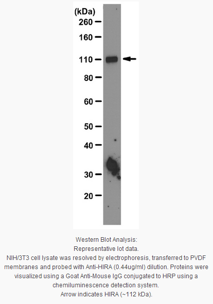
Evaluated by Western Blot in NIH/3T3 cell lysate.
Western Blot Analysis: 0.44 µg/ml of this antibody detected HIRA on 10 µg of NIH/3T3 cell lysate.
Usage Statement
Unless otherwise stated in our catalog or other company documentation accompanying the product(s), our products are intended for research use only and are not to be used for any other purpose, which includes but is not limited to, unauthorized commercial uses, in vitro diagnostic uses, ex vivo or in vivo therapeutic uses or any type of consumption or application to humans or animals.
Storage and Shipping Information
Storage Conditions
Stable for 1 year at 2-8°C from date of receipt.
Packaging Information
Material Size
100 µg
原厂资料:
Key Spec Table
Species Reactivity
Key Applications
Host
Format
Antibody Type
H, M
IP, WB
M
Purified
Monoclonal Antibody
Description
Catalogue Number
04-1488
Description
Anti-HIRA Antibody, clone WC119
Alternate Names
TUP1-like enhancer of split protein 1
DiGeorge critical region gene 1
HIR (histone cell cycle regulation defective) homolog A (S. cerevisiae)
HIR histone cell cycle regulation defective homolog A
HIR histone cell cycle regulation defective homolog A (S. cerevisiae)
Background Information
HIRA (TUP1 like enhancer of split protein 1) is a protein localized primarily in the nucleus. It functions as a histone chaparone to preferentially place histone H3.3 on nucleosome. It is broadly expressed in a variety of tissues and is expressed during embryogenesis. Data suggests HIRA plays a part in mechanisms of transcriptional regulation similar to that played by yeast HIR1 and HIR2 together. The HIRA protein has been shown to interact with HIRIP3, HIRIP5, histones H2B and H4, and PAX3. HIRA may play a part in the etiology of the DiGeorge syndrome (DGS), a developmental disorder. The clinical features of this disease include absence or hypoplasia of the thymus and parathyroid glands, cardiovascular malformations, facial dysplasia, a cleft palate and mental retardation.
Product Information
Format
Purified
Control
NIH/3T3 cell lysate
Presentation
Purified mouse monoclonal IgG1κ in buffer containing 0.1 M Tris-Glycine (pH 7.4, 150 mM NaCl) with 0.05% sodium azide.
Applications
Application
Anti-HIRA Antibody, clone WC119 is a Mouse Monoclonal Antibody for detection of HIRA also known as HIR (histone cell cycle regulation defective) homolog & has been validated in WB & IP.
Key Applications
Immunoprecipitation
Western Blotting
Application Notes
Immunoprecipitation Analysis: A previous lot was used by an independent laboratory in IP. (Hall, C., et al. (2001). Molecular and Cellular Biology. 21(5):1854-1865.)
Biological Information
Immunogen
GST-tagged recombinant protein corresponding to human HIRA.
Epitope
Unknown
Clone
WC119
Concentration
Please refer to the Certificate of Analysis for the lot-specific concentration.
Host
Mouse
Specificity
This antibody recognizes HIRA.
Isotype
IgG1κ
Species Reactivity
Human Mouse
Species Reactivity Note
Demonstrated to react with mouse. Predicted to react with human based on 100% sequence homology.
This gene encodes a histone chaperone that preferentially places the variant histone H3.3 in nucleosomes. Orthologs of this gene in yeast, flies, and plants are necessary for the formation of transcriptionally silent heterochomatin. This gene plays an important role in the formation of the senescence-associated heterochromatin foci. These foci likely mediate the irreversible cell cycle changes that occur in senescent cells. It is considered the primary candidate gene in some haploinsufficiency syndromes such as DiGeorge syndrome, and insufficient production of the gene may disrupt normal embryonic development. [provided by RefSeq].
FUNCTION: Cooperates with ASF1A to promote replication-independent chromatin assembly. Required for the periodic repression of histone gene transcription during the cell cycle. Required for the formation of senescence-associated heterochromatin foci (SAHF) and efficient senescence-associated cell cycle exit.
SUBUNIT STRUCTURE: Interacts with histone H3F3B, PAX3 and PAX7 By similarity. Interacts with CCNA1, HIRIP3, NFU1/HIRIP5 and histone H2B. Part of a complex which includes ASF1A, CABIN1, histone H3.3, histone H4 and UBN1.
SUBCELLULAR LOCATION: Nucleus. Note: Primarily, though not exclusively, localized to the nucleus. Localizes to PML bodies immediately prior to onset of senescence.
TISSUE SPECIFICTY: Expressed at high levels in kidney, pancreas and skeletal muscle and at lower levels in brain, heart, liver, lung, and placenta. Ref.6
DEVELOPMENTAL STAGE: Expressed during embryogenesis.
PTM: Sumoylated.
Phosphorylated by CDK2/CCNA1 and CDK2/CCNE1 on Thr-555 in vitro. Also phosphorylated on Thr-555 and Ser-687 in vivo.
INVOLVEMENT IN DISEASE: May play a part in the etiology of the DiGeorge syndrome (DGS), a developmental disorder due to an abnormal development of the third and fourth pharyngeal pouches. The clinical features include absence or hypoplasia of the thymus and parathyroid glands, cardiovascular malformations, facial dysplasia, a cleft palate and mental retardation.
SEQUENCE SIMILARITIES: Belongs to the WD repeat HIR1 family.
Contains 8 WD repeats.
Product Usage Statements
Quality Assurance
Evaluated by Western Blot in NIH/3T3 cell lysate.
Western Blot Analysis: 0.44 µg/ml of this antibody detected HIRA on 10 µg of NIH/3T3 cell lysate.
Usage Statement
Unless otherwise stated in our catalog or other company documentation accompanying the product(s), our products are intended for research use only and are not to be used for any other purpose, which includes but is not limited to, unauthorized commercial uses, in vitro diagnostic uses, ex vivo or in vivo therapeutic uses or any type of consumption or application to humans or animals.


 京公网安备11010802025653 版权所有:北京逸优科技有限公司
