Lymphoid Enhancer Factor-1 (LEF-1) is an HMG 1/2-like DNA binding/bending protein and is a member of the LEF/TCF transcription factor family. There are four LEF/TCF family members in mammalian systems (LEF-1, TCF-1, TCF-3 and TCF-4), and orthologs to these factors have been identified in many different species. LEF/TCFs are downstream mediators of Wnt/Wingless signals. Wnt signaling drives cell polarity, cell fate, and cell growth decisions in embryonic tissues and in post-natal tissues that continue to develop from mitotically active stem cell precursors. Misregulation of Wnt signaling is also implicated as a root cause of many different cancers, such as colon cancer, melanoma, breast cancer, prostate cancer and others. This antibody recognizes an epitope in amino acids 236-242 of LEF-1. This region is found within the transactivation domain of LEF-1. However, it is not present in some splice variants.
Product Information
Format
Unpurified
Control
Jurkat cell lysate
Presentation
Rabbit Monoclonal in buffer containing 50 mM Tris-Glycine (pH 7.4), 0.15 M NaCl containing 40% Glycerol, 0.01% sodium azide and 0.05% BSA.
Applications
Application
Please note that this product will not be available for sale after March 15, 2015. Please select one of the other antibodies against this target.
Key Applications
Flow Cytometry
Western Blotting
Immunocytochemistry
Immunohistochemistry
Application Notes
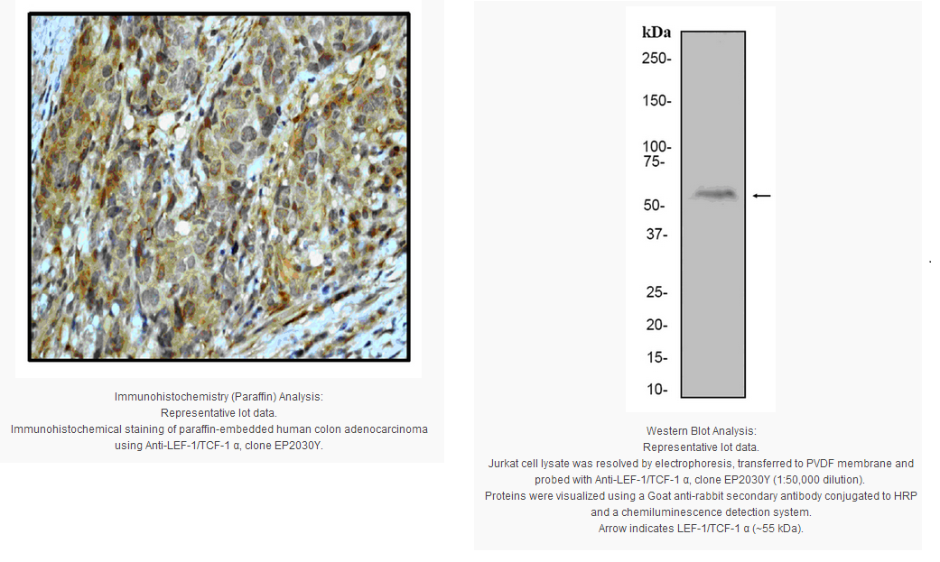
Immunohistochemistry Analysis: A 1:50 dilution from a representative lot detected LEF-1/TCF-1 α in human colon adenocarcinoma tissue.
Immunocytochemistry Analysis: A 1:100-250 dilution from a representative lot was used in IC.
Flow Cytometry: A 1:20 dilution of a representative lot was used in flow cytometry.
Biological Information
Immunogen
Synthetic peptide corresponding to human LEF-1/TCF-1 α.
Clone
EP2030Y
Host
Rabbit
Specificity
This antibody recognizes LEF-1/TCF-1 α. Recognition mpas to an epitope in amino acids 236-242 of LEF-1. This region is found within the transactivation domain of LEF-1. However, it is not present in some splice variants.
Evaluated by Western Blot on Jurkat cell lysates.
Western Blot Analysis: A 1:25,000-50,000 dilution of this antibody was used to detect LEF-1/TCF-1 α in Jurkat cell lysate.
Usage Statement
Unless otherwise stated in our catalog or other company documentation accompanying the product(s), our products are intended for research use only and are not to be used for any other purpose, which includes but is not limited to, unauthorized commercial uses, in vitro diagnostic uses, ex vivo or in vivo therapeutic uses or any type of consumption or application to humans or animals.
Storage and Shipping Information
Storage Conditions
Stable for 1 year at -20ºC from date of receipt.
Handling Recommendations: Upon first thaw, and prior to removing the cap, centrifuge the vial and gently mix the solution. Aliquot into microcentrifuge tubes and store at -20°C. Avoid repeated freeze/thaw cycles, which may damage IgG and affect product performance. Note: Variability in freezer temperatures below -20°C may cause glycerol containing solutions to become frozen during storage.
Lymphoid Enhancer Factor-1 (LEF-1) is an HMG 1/2-like DNA binding/bending protein and is a member of the LEF/TCF transcription factor family. There are four LEF/TCF family members in mammalian systems (LEF-1, TCF-1, TCF-3 and TCF-4), and orthologs to these factors have been identified in many different species. LEF/TCFs are downstream mediators of Wnt/Wingless signals. Wnt signaling drives cell polarity, cell fate, and cell growth decisions in embryonic tissues and in post-natal tissues that continue to develop from mitotically active stem cell precursors. Misregulation of Wnt signaling is also implicated as a root cause of many different cancers, such as colon cancer, melanoma, breast cancer, prostate cancer and others. This antibody recognizes an epitope in amino acids 236-242 of LEF-1. This region is found within the transactivation domain of LEF-1. However, it is not present in some splice variants.
Product Information
Format
Unpurified
Control
Jurkat cell lysate
Presentation
Rabbit Monoclonal in buffer containing 50 mM Tris-Glycine (pH 7.4), 0.15 M NaCl containing 40% Glycerol, 0.01% sodium azide and 0.05% BSA.
Applications
Application
Please note that this product will not be available for sale after March 15, 2015. Please select one of the other antibodies against this target.
Key Applications
Flow Cytometry
Western Blotting
Immunocytochemistry
Immunohistochemistry
Application Notes
Immunohistochemistry Analysis: A 1:50 dilution from a representative lot detected LEF-1/TCF-1 α in human colon adenocarcinoma tissue.
Immunocytochemistry Analysis: A 1:100-250 dilution from a representative lot was used in IC.
Flow Cytometry: A 1:20 dilution of a representative lot was used in flow cytometry.
Biological Information
Immunogen
Synthetic peptide corresponding to human LEF-1/TCF-1 α.
Clone
EP2030Y
Host
Rabbit
Specificity
This antibody recognizes LEF-1/TCF-1 α. Recognition mpas to an epitope in amino acids 236-242 of LEF-1. This region is found within the transactivation domain of LEF-1. However, it is not present in some splice variants.
Evaluated by Western Blot on Jurkat cell lysates.
Western Blot Analysis: A 1:25,000-50,000 dilution of this antibody was used to detect LEF-1/TCF-1 α in Jurkat cell lysate.
Usage Statement
Unless otherwise stated in our catalog or other company documentation accompanying the product(s), our products are intended for research use only and are not to be used for any other purpose, which includes but is not limited to, unauthorized commercial uses, in vitro diagnostic uses, ex vivo or in vivo therapeutic uses or any type of consumption or application to humans or animals.
Storage and Shipping Information
Storage Conditions
Stable for 1 year at -20ºC from date of receipt.
Handling Recommendations: Upon first thaw, and prior to removing the cap, centrifuge the vial and gently mix the solution. Aliquot into microcentrifuge tubes and store at -20°C. Avoid repeated freeze/thaw cycles, which may damage IgG and affect product performance. Note: Variability in freezer temperatures below -20°C may cause glycerol containing solutions to become frozen during storage.


 京公网安备11010802025653 版权所有:北京逸优科技有限公司
