FADD was originally isolated as a protein that bound to the cytoplasmic domain of Fas in the yeast two-hybrid system. Sequence analysis revealed a region homologous to the death domain of Fas and TNFR-1. Subsequent biochemical studies have shown that FADD associates with FAS through interaction of the death domains. When over expressed in several cell lines, FADD induces apoptosis, which can be blocked by CrmA, an inhibitor of inerleukin-1-beta-converting enzyme. This evidence suggests that FADD plays a role in Fas-mediated apoptosis.
Product Information
Format
Unpurified
Control
Jurkat lysates
Presentation
Rabbit Monoclonal in buffer containing 50 mM Tris-Glycine (pH 7.4), 0.18 M NaCl containing 40% Glycerol, 0.25% sodium azide, 0.25 mM EDTA, 25 mM Citric Acid, and 0.5% BSA.
Applications
Application
Please note that this product will not be available for sale after March 15, 2015. Please select one of the other antibodies against this target. This Anti-FADD (N-terminal) Antibody, clone EP887Y, Rabbit is validated for use in IH(P), WB for the detection of FADD (N-terminal).
Key Applications
Western Blotting
Immunohistochemistry (Paraffin)
Application Notes
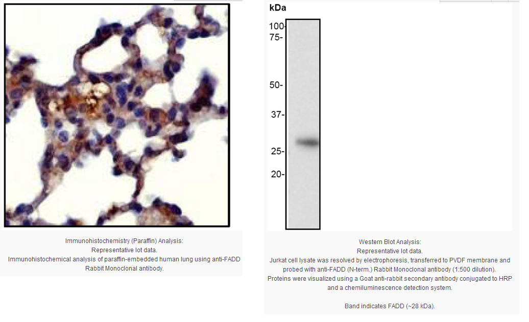
Immunohistochemistry Analysis: 1:250 dilution from a previous lot detected FADD in human lung tissue.
Biological Information
Immunogen
Synthetic peptide corresponding to residues near the N-terminus of human FADD.
FUNCTION:Apoptotic adaptor molecule that recruits caspase-8 or caspase-10 to the activated Fas (CD95) or TNFR-1 receptors. The resulting aggregate called the death-inducing signaling complex (DISC) performs caspase-8 proteolytic activation. Active caspase-8 initiates the subsequent cascade of caspases mediating apoptosis.
SUBUNIT STRUCTURE:Interacts with CFLAR, PEA15 and MBD4. When phosphorylated, part of a complex containing HIPK3 and FAS. May interact with MAVS/IPS1. Interacts with MOCV v-CFLAR protein and LRDD.
TISSUE SPECIFICITY:Expressed in a wide variety of tissues, except for peripheral blood mononuclear leukocytes.
DOMAIN:Contains a death domain involved in the binding of the corresponding domain within Fas receptor.
PTM:Phosphorylated.
SEQUENCE SIMILARITY:Contains 1 death domain.
Contains 1 DED (death effector) domain.
Product Usage Statements
Quality Assurance
Evaluated by Western Blot on Jurkat cell lysates.
Western Blotting Analysis:
1:500 dilution of this antibody was used to detect FADD in Jurkat cell lysate.
Usage Statement
Unless otherwise stated in our catalog or other company documentation accompanying the product(s), our products are intended for research use only and are not to be used for any other purpose, which includes but is not limited to, unauthorized commercial uses, in vitro diagnostic uses, ex vivo or in vivo therapeutic uses or any type of consumption or application to humans or animals.
Storage and Shipping Information
Storage Conditions
Stable for 1 year at -20ºC from date of receipt.
Handling Recommendations: Upon receipt, and prior to removing the cap, centrifuge the vial and gently mix the solution. Aliquot into microcentrifuge tubes and store at -20°C. Avoid repeated freeze/thaw cycles, which may damage IgG and affect product performance. Note: Variability in freezer temperatures below -20°C may cause glycerol containing solutions to become frozen during storage.
FADD was originally isolated as a protein that bound to the cytoplasmic domain of Fas in the yeast two-hybrid system. Sequence analysis revealed a region homologous to the death domain of Fas and TNFR-1. Subsequent biochemical studies have shown that FADD associates with FAS through interaction of the death domains. When over expressed in several cell lines, FADD induces apoptosis, which can be blocked by CrmA, an inhibitor of inerleukin-1-beta-converting enzyme. This evidence suggests that FADD plays a role in Fas-mediated apoptosis.
Product Information
Format
Unpurified
Control
Jurkat lysates
Presentation
Rabbit Monoclonal in buffer containing 50 mM Tris-Glycine (pH 7.4), 0.18 M NaCl containing 40% Glycerol, 0.25% sodium azide, 0.25 mM EDTA, 25 mM Citric Acid, and 0.5% BSA.
Applications
Application
Please note that this product will not be available for sale after March 15, 2015. Please select one of the other antibodies against this target. This Anti-FADD (N-terminal) Antibody, clone EP887Y, Rabbit is validated for use in IH(P), WB for the detection of FADD (N-terminal).
Key Applications
Western Blotting
Immunohistochemistry (Paraffin)
Application Notes
Immunohistochemistry Analysis: 1:250 dilution from a previous lot detected FADD in human lung tissue.
Biological Information
Immunogen
Synthetic peptide corresponding to residues near the N-terminus of human FADD.
FUNCTION:Apoptotic adaptor molecule that recruits caspase-8 or caspase-10 to the activated Fas (CD95) or TNFR-1 receptors. The resulting aggregate called the death-inducing signaling complex (DISC) performs caspase-8 proteolytic activation. Active caspase-8 initiates the subsequent cascade of caspases mediating apoptosis.
SUBUNIT STRUCTURE:Interacts with CFLAR, PEA15 and MBD4. When phosphorylated, part of a complex containing HIPK3 and FAS. May interact with MAVS/IPS1. Interacts with MOCV v-CFLAR protein and LRDD.
TISSUE SPECIFICITY:Expressed in a wide variety of tissues, except for peripheral blood mononuclear leukocytes.
DOMAIN:Contains a death domain involved in the binding of the corresponding domain within Fas receptor.
PTM:Phosphorylated.
SEQUENCE SIMILARITY:Contains 1 death domain.
Contains 1 DED (death effector) domain.
Product Usage Statements
Quality Assurance
Evaluated by Western Blot on Jurkat cell lysates.
Western Blotting Analysis:
1:500 dilution of this antibody was used to detect FADD in Jurkat cell lysate.
Usage Statement
Unless otherwise stated in our catalog or other company documentation accompanying the product(s), our products are intended for research use only and are not to be used for any other purpose, which includes but is not limited to, unauthorized commercial uses, in vitro diagnostic uses, ex vivo or in vivo therapeutic uses or any type of consumption or application to humans or animals.
Storage and Shipping Information
Storage Conditions
Stable for 1 year at -20ºC from date of receipt.
Handling Recommendations: Upon receipt, and prior to removing the cap, centrifuge the vial and gently mix the solution. Aliquot into microcentrifuge tubes and store at -20°C. Avoid repeated freeze/thaw cycles, which may damage IgG and affect product performance. Note: Variability in freezer temperatures below -20°C may cause glycerol containing solutions to become frozen during storage.


 京公网安备11010802025653 版权所有:北京逸优科技有限公司
