Neurofilaments are a type of intermediate filament that serve as major elements of the cytoskeleton supporting the axon cytoplasm. They are the most abundant fibrillar components of the axon, being on average 3-10 times more frequent than axonal microtubules. Neurofilaments (10nm in dia.) are built from three intertwined protofibrils which are themselves composed of two tetrameric protofilament complex of monomeric proteins. The neurofilament triplet proteins (68/70, 160, and 200 kDa) occur in both the central and peripheral nervous system and are usually neuron specific. The 68/70 kDa NF-L protein can self-assemble into a filamentous structure, however the 160 kDa NF-M and 200 kDa NF-H proteins require the presence of the 68/70 kDa NF-L protein to co-assemble. Neuromas, ganglioneuromas, gangliogliomas, ganglioneuroblastomas and neuroblastomas stain positively for neurofilaments. Although typically restricted to neurons, neurofilaments have been detected in paragangliomas and adrenal and extra-adrenal pheochromocytomas. Carcinoids, neuroendocrine carcinomas of the skin, and oat cell carcinomas of the lung also express neurofilaments.
Product Information
Format
Unpurified
Control
SHSY5Y cell lysate
Presentation
Rabbit Monoclonal in buffer containing 50 mM Tris-Glycine (pH 7.4), 0.15 M NaCl containing 40% Glycerol, 0.01% sodium azide and 0.05% BSA.
Applications
Application
Please note that this product will not be available for sale after March 15, 2015. Please select one of the other antibodies against this target.
Key Applications
Immunoprecipitation
Western Blotting
Flow Cytometry
Application Notes
Flow Cytometry: A 1:70 dilution of a previous lot was used in flow cytometry.
Immunoprecipitation Analysis: A 1:50 dilution from a previous lot was used in IP.
Biological Information
Immunogen
Synthetic peptide corresponding to residues near the C-terminus of human Neurofilament-L.
Epitope
C-terminus
Clone
EP675Y
Host
Rabbit
Specificity
This antibody recognizes Neurofilament-L at and around the C-terminus.
Neurofilaments are type IV intermediate filament heteropolymers composed of light, medium, and heavy chains. Neurofilaments comprise the axoskeleton and they functionally maintain the neuronal caliber. They may also play a role in intracellular transport to axons and dendrites. This gene encodes the light chain neurofilament protein. Mutations in this gene cause Charcot-Marie-Tooth disease types 1F (CMT1F) and 2E (CMT2E), disorders of the peripheral nervous system that are characterized by distinct neuropathies. A pseudogene has been identified on chromosome Y.
FUNCTION:Neurofilaments usually contain three intermediate filament proteins: L, M, and H which are involved in the maintenance of neuronal caliber. SUBUNIT STRUCTURE:Interacts with RGNEF By similarity. DOMAIN:The extra mass and high charge density that distinguish the neurofilament proteins from all other intermediate filament proteins are due to the tailpiece extensions. This region may form a charged scaffolding structure suitable for interaction with other neuronal components or ions. PTM:O-glycosylated By similarity. INVOLVEMENT IN DISEASE:Defects in NEFL are the cause of Charcot-Marie-Tooth disease type 1F (CMT1F) [MIM:607734]. CMT1F is a form of Charcot-Marie-Tooth disease, the most common inherited disorder of the peripheral nervous system. Charcot-Marie-Tooth disease is classified in two main groups on the basis of electrophysiologic properties and histopathology: primary peripheral demyelinating neuropathy or CMT1, and primary peripheral axonal neuropathy or CMT2. Neuropathies of the CMT1 group are characterized by severely reduced nerve conduction velocities (less than 38 m/sec), segmental demyelination and remyelination with onion bulb formations on nerve biopsy, slowly progressive distal muscle atrophy and weakness, absent deep tendon reflexes, and hollow feet. CMT1F is characterized by onset in infancy or childhood (range 1 to 13 years). Defects in NEFL are the cause of Charcot-Marie-Tooth disease type 2E (CMT2E) [MIM:607684]. CMT2E is an autosomal dominant form of Charcot-Marie-Tooth disease type 2. Neuropathies of the CMT2 group are characterized by signs of axonal regeneration in the absence of obvious myelin alterations, normal or slightly reduced nerve conduction velocities, and progressive distal muscle weakness and atrophy. MISCELLANEOUS:NF-L is the most abundant of the three neurofilament proteins and, as the other nonepithelial intermediate filament proteins, it can form homopolymeric 10-nm filaments. SEQUENCE SIMILARITIES:Belongs to the intermediate filament family.
Product Usage Statements
Quality Assurance
Evaluated by Western Blot on SHSY5Y cell lysates.
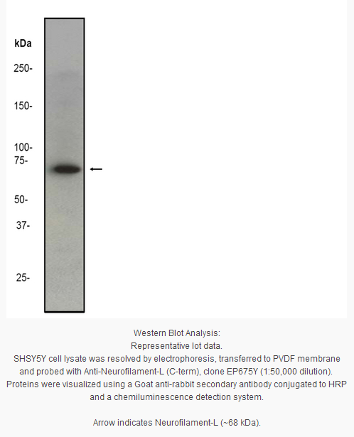
Western Blot Analysis: 1:50,000 dilution of this antibody was used to detect Neurofilament L in SHSY5Y cell lysate.
Usage Statement
Unless otherwise stated in our catalog or other company documentation accompanying the product(s), our products are intended for research use only and are not to be used for any other purpose, which includes but is not limited to, unauthorized commercial uses, in vitro diagnostic uses, ex vivo or in vivo therapeutic uses or any type of consumption or application to humans or animals.
Storage and Shipping Information
Storage Conditions
Stable for 1 year at -20ºC from date of receipt.
Handling Recommendations: Upon first thaw, and prior to removing the cap, centrifuge the vial and gently mix the solution. Aliquot into microcentrifuge tubes and store at -20°C. Avoid repeated freeze/thaw cycles, which may damage IgG and affect product performance. Note: Variability in freezer temperatures below -20°C may cause glycerol containing solutions to become frozen during storage.
Neurofilaments are a type of intermediate filament that serve as major elements of the cytoskeleton supporting the axon cytoplasm. They are the most abundant fibrillar components of the axon, being on average 3-10 times more frequent than axonal microtubules. Neurofilaments (10nm in dia.) are built from three intertwined protofibrils which are themselves composed of two tetrameric protofilament complex of monomeric proteins. The neurofilament triplet proteins (68/70, 160, and 200 kDa) occur in both the central and peripheral nervous system and are usually neuron specific. The 68/70 kDa NF-L protein can self-assemble into a filamentous structure, however the 160 kDa NF-M and 200 kDa NF-H proteins require the presence of the 68/70 kDa NF-L protein to co-assemble. Neuromas, ganglioneuromas, gangliogliomas, ganglioneuroblastomas and neuroblastomas stain positively for neurofilaments. Although typically restricted to neurons, neurofilaments have been detected in paragangliomas and adrenal and extra-adrenal pheochromocytomas. Carcinoids, neuroendocrine carcinomas of the skin, and oat cell carcinomas of the lung also express neurofilaments.
Product Information
Format
Unpurified
Control
SHSY5Y cell lysate
Presentation
Rabbit Monoclonal in buffer containing 50 mM Tris-Glycine (pH 7.4), 0.15 M NaCl containing 40% Glycerol, 0.01% sodium azide and 0.05% BSA.
Applications
Application
Please note that this product will not be available for sale after March 15, 2015. Please select one of the other antibodies against this target.
Key Applications
Immunoprecipitation
Western Blotting
Flow Cytometry
Application Notes
Flow Cytometry: A 1:70 dilution of a previous lot was used in flow cytometry.
Immunoprecipitation Analysis: A 1:50 dilution from a previous lot was used in IP.
Biological Information
Immunogen
Synthetic peptide corresponding to residues near the C-terminus of human Neurofilament-L.
Epitope
C-terminus
Clone
EP675Y
Host
Rabbit
Specificity
This antibody recognizes Neurofilament-L at and around the C-terminus.
Neurofilaments are type IV intermediate filament heteropolymers composed of light, medium, and heavy chains. Neurofilaments comprise the axoskeleton and they functionally maintain the neuronal caliber. They may also play a role in intracellular transport to axons and dendrites. This gene encodes the light chain neurofilament protein. Mutations in this gene cause Charcot-Marie-Tooth disease types 1F (CMT1F) and 2E (CMT2E), disorders of the peripheral nervous system that are characterized by distinct neuropathies. A pseudogene has been identified on chromosome Y.
FUNCTION:Neurofilaments usually contain three intermediate filament proteins: L, M, and H which are involved in the maintenance of neuronal caliber. SUBUNIT STRUCTURE:Interacts with RGNEF By similarity. DOMAIN:The extra mass and high charge density that distinguish the neurofilament proteins from all other intermediate filament proteins are due to the tailpiece extensions. This region may form a charged scaffolding structure suitable for interaction with other neuronal components or ions. PTM:O-glycosylated By similarity. INVOLVEMENT IN DISEASE:Defects in NEFL are the cause of Charcot-Marie-Tooth disease type 1F (CMT1F) [MIM:607734]. CMT1F is a form of Charcot-Marie-Tooth disease, the most common inherited disorder of the peripheral nervous system. Charcot-Marie-Tooth disease is classified in two main groups on the basis of electrophysiologic properties and histopathology: primary peripheral demyelinating neuropathy or CMT1, and primary peripheral axonal neuropathy or CMT2. Neuropathies of the CMT1 group are characterized by severely reduced nerve conduction velocities (less than 38 m/sec), segmental demyelination and remyelination with onion bulb formations on nerve biopsy, slowly progressive distal muscle atrophy and weakness, absent deep tendon reflexes, and hollow feet. CMT1F is characterized by onset in infancy or childhood (range 1 to 13 years). Defects in NEFL are the cause of Charcot-Marie-Tooth disease type 2E (CMT2E) [MIM:607684]. CMT2E is an autosomal dominant form of Charcot-Marie-Tooth disease type 2. Neuropathies of the CMT2 group are characterized by signs of axonal regeneration in the absence of obvious myelin alterations, normal or slightly reduced nerve conduction velocities, and progressive distal muscle weakness and atrophy. MISCELLANEOUS:NF-L is the most abundant of the three neurofilament proteins and, as the other nonepithelial intermediate filament proteins, it can form homopolymeric 10-nm filaments. SEQUENCE SIMILARITIES:Belongs to the intermediate filament family.
Product Usage Statements
Quality Assurance
Evaluated by Western Blot on SHSY5Y cell lysates.
Western Blot Analysis: 1:50,000 dilution of this antibody was used to detect Neurofilament L in SHSY5Y cell lysate.
Usage Statement
Unless otherwise stated in our catalog or other company documentation accompanying the product(s), our products are intended for research use only and are not to be used for any other purpose, which includes but is not limited to, unauthorized commercial uses, in vitro diagnostic uses, ex vivo or in vivo therapeutic uses or any type of consumption or application to humans or animals.
Storage and Shipping Information
Storage Conditions
Stable for 1 year at -20ºC from date of receipt.
Handling Recommendations: Upon first thaw, and prior to removing the cap, centrifuge the vial and gently mix the solution. Aliquot into microcentrifuge tubes and store at -20°C. Avoid repeated freeze/thaw cycles, which may damage IgG and affect product performance. Note: Variability in freezer temperatures below -20°C may cause glycerol containing solutions to become frozen during storage.


 京公网安备11010802025653 版权所有:北京逸优科技有限公司
