Glial fibrillary acidic protein is a class-III intermediate filament. GFAP is the main constituent of intermediate filaments in astrocytes and serves as a cell specific marker that distinguishes differentiated astrocytes from other glial cells during the development of the central nervous system.
Product Information
Format
Unpurified
Control
Human and Rat brain lysates
Presentation
Rabbit Monoclonal in buffer containing 50 mM Tris-Glycine (pH 7.4), 0.15 M NaCl containing 40% Glycerol, 0.01% sodium azide and 0.05% BSA.
Applications
Application
Please note that this product will not be available for sale after March 15, 2015. Please select one of the other antibodies against this target. Detect GFAP (N-term) using this Anti-GFAP (N-term) Antibody, clone EPR1034Y, Rabbit validated for use in WB, IH(P), IC & IP.
Key Applications
Immunoprecipitation
Western Blotting
Immunohistochemistry (Paraffin)
Immunocytochemistry
Application Notes
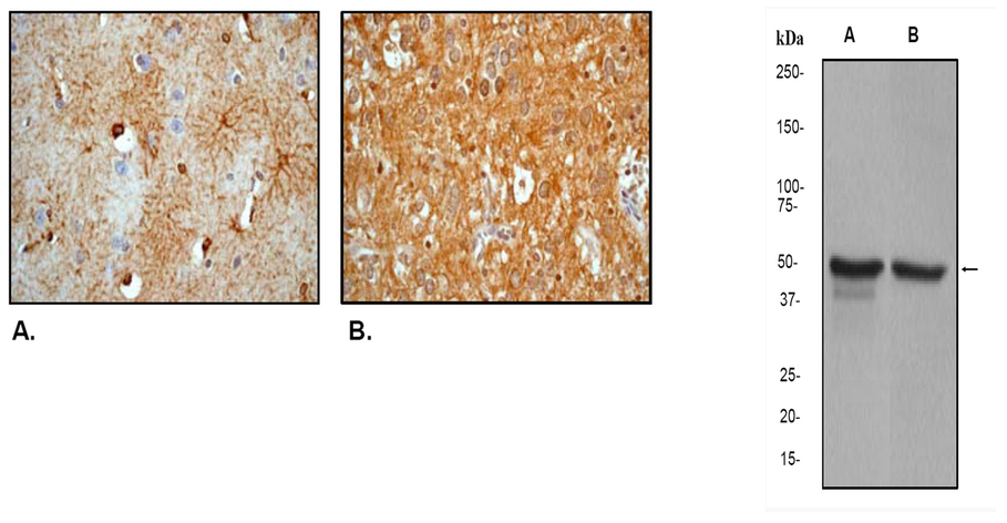
Immunohistochemistry Analysis: A 1:250-500 dilution from a previous lot detected GFAP in human brain and human glioma tissue.
Immunocytochemistry Analysis: A 1:100-250 dilution from a previous lot was used in IC.
Immunoprecipitation Analysis: A 1:40 dilution from a previous lot was used in IP.
Biological Information
Immunogen
Synthetic peptide corresponding to residues in the N-terminus of human GFAP.
Epitope
N-terminus
Clone
EPR1034Y
Host
Rabbit
Specificity
This antibody recognizes GFAP at and around the N-terminus.
This gene encodes one of the major intermediate filament proteins of mature astrocytes. It is used as a marker to distinguish astrocytes from other glial cells during development. Mutations in this gene cause Alexander disease, a rare disorder of astrocytes in the central nervous system. Alternative splicing results in multiple transcript variants encoding distinct isoforms.
FUNCTION:GFAP, a class-III intermediate filament, is a cell-specific marker that, during the development of the central nervous system, distinguishes astrocytes from other glial cells. SUBUNIT STRUCTURE:Interacts with SYNM By similarity. Isoform 3 interacts with PSEN1 (via N-terminus). SUBCELLULAR LOCATION:Cytoplasm. Note: Associated with intermediate filaments. TISSUE SPECIFICITY:Expressed in cells lacking fibronectin. INVOLVEMENT IN DISEASE:Defects in GFAP are a cause of Alexander disease (ALEXD) [MIM:203450]. Alexander disease is a rare disorder of the central nervous system. It is a progressive leukoencephalopathy whose hallmark is the widespread accumulation of Rosenthal fibers which are cytoplasmic inclusions in astrocytes. The most common form affects infants and young children, and is characterized by progressive failure of central myelination, usually leading to death usually within the first decade. Infants with Alexander disease develop a leukoencephalopathy with macrocephaly, seizures, and psychomotor retardation. Patients with juvenile or adult forms typically experience ataxia, bulbar signs and spasticity, and a more slowly progressive course. SEQUENCE SIMILARITIES:Belongs to the intermediate filament family.
Product Usage Statements
Quality Assurance
Evaluated by Western Blot on Human and Rat brain cell lysates.
Western Blot Analysis: 1:50,000-100,000 dilution of this antibody was used to detect GFAP in human and rat brain cell lysates.
Usage Statement
Unless otherwise stated in our catalog or other company documentation accompanying the product(s), our products are intended for research use only and are not to be used for any other purpose, which includes but is not limited to, unauthorized commercial uses, in vitro diagnostic uses, ex vivo or in vivo therapeutic uses or any type of consumption or application to humans or animals.
Storage and Shipping Information
Storage Conditions
Stable for 1 year at -20ºC from date of receipt.
Handling Recommendations: Upon first thaw, and prior to removing the cap, centrifuge the vial and gently mix the solution. Aliquot into microcentrifuge tubes and store at -20°C. Avoid repeated freeze/thaw cycles, which may damage IgG and affect product performance. Note: Variability in freezer temperatures below -20°C may cause glycerol containing solutions to become frozen during storage.
Glial fibrillary acidic protein is a class-III intermediate filament. GFAP is the main constituent of intermediate filaments in astrocytes and serves as a cell specific marker that distinguishes differentiated astrocytes from other glial cells during the development of the central nervous system.
Product Information
Format
Unpurified
Control
Human and Rat brain lysates
Presentation
Rabbit Monoclonal in buffer containing 50 mM Tris-Glycine (pH 7.4), 0.15 M NaCl containing 40% Glycerol, 0.01% sodium azide and 0.05% BSA.
Applications
Application
Please note that this product will not be available for sale after March 15, 2015. Please select one of the other antibodies against this target. Detect GFAP (N-term) using this Anti-GFAP (N-term) Antibody, clone EPR1034Y, Rabbit validated for use in WB, IH(P), IC & IP.
Key Applications
Immunoprecipitation
Western Blotting
Immunohistochemistry (Paraffin)
Immunocytochemistry
Application Notes
Immunohistochemistry Analysis: A 1:250-500 dilution from a previous lot detected GFAP in human brain and human glioma tissue.
Immunocytochemistry Analysis: A 1:100-250 dilution from a previous lot was used in IC.
Immunoprecipitation Analysis: A 1:40 dilution from a previous lot was used in IP.
Biological Information
Immunogen
Synthetic peptide corresponding to residues in the N-terminus of human GFAP.
Epitope
N-terminus
Clone
EPR1034Y
Host
Rabbit
Specificity
This antibody recognizes GFAP at and around the N-terminus.
This gene encodes one of the major intermediate filament proteins of mature astrocytes. It is used as a marker to distinguish astrocytes from other glial cells during development. Mutations in this gene cause Alexander disease, a rare disorder of astrocytes in the central nervous system. Alternative splicing results in multiple transcript variants encoding distinct isoforms.
FUNCTION:GFAP, a class-III intermediate filament, is a cell-specific marker that, during the development of the central nervous system, distinguishes astrocytes from other glial cells. SUBUNIT STRUCTURE:Interacts with SYNM By similarity. Isoform 3 interacts with PSEN1 (via N-terminus). SUBCELLULAR LOCATION:Cytoplasm. Note: Associated with intermediate filaments. TISSUE SPECIFICITY:Expressed in cells lacking fibronectin. INVOLVEMENT IN DISEASE:Defects in GFAP are a cause of Alexander disease (ALEXD) [MIM:203450]. Alexander disease is a rare disorder of the central nervous system. It is a progressive leukoencephalopathy whose hallmark is the widespread accumulation of Rosenthal fibers which are cytoplasmic inclusions in astrocytes. The most common form affects infants and young children, and is characterized by progressive failure of central myelination, usually leading to death usually within the first decade. Infants with Alexander disease develop a leukoencephalopathy with macrocephaly, seizures, and psychomotor retardation. Patients with juvenile or adult forms typically experience ataxia, bulbar signs and spasticity, and a more slowly progressive course. SEQUENCE SIMILARITIES:Belongs to the intermediate filament family.
Product Usage Statements
Quality Assurance
Evaluated by Western Blot on Human and Rat brain cell lysates.
Western Blot Analysis: 1:50,000-100,000 dilution of this antibody was used to detect GFAP in human and rat brain cell lysates.
Usage Statement
Unless otherwise stated in our catalog or other company documentation accompanying the product(s), our products are intended for research use only and are not to be used for any other purpose, which includes but is not limited to, unauthorized commercial uses, in vitro diagnostic uses, ex vivo or in vivo therapeutic uses or any type of consumption or application to humans or animals.
Storage and Shipping Information
Storage Conditions
Stable for 1 year at -20ºC from date of receipt.
Handling Recommendations: Upon first thaw, and prior to removing the cap, centrifuge the vial and gently mix the solution. Aliquot into microcentrifuge tubes and store at -20°C. Avoid repeated freeze/thaw cycles, which may damage IgG and affect product performance. Note: Variability in freezer temperatures below -20°C may cause glycerol containing solutions to become frozen during storage.


 京公网安备11010802025653 版权所有:北京逸优科技有限公司
