STAT proteins (Signal Transduction and Activators of Transcription) are latent cytoplasmic transcription factors that have the dual function of signal transduction and activation of transcription. STATs are activated by tyrosine phosphorylation in response to different ligands, after which they translocate to the cell nucleus. The N-terminal region is highly homologous among the STAT proteins and surrounds a completely conserved arginine residue. STATs are a part of the JAK-STAT signaling pathway – a major pathway of the immune system. All cytokines transduce critical signals through this pathway. STAT3 has been shown to be activated by IFN alpha but not IFN beta. The transcription factors associated with STAT3 are cJun and cyclic AMP responsive enhancer binding protein (CREB). Deletion of the STAT3 gene in knock out mice was lethal at the early embryonic stage.
Product Information
Format
Unpurified
Control
HeLa cell lysate
Presentation
Rabbit Monoclonal in buffer containing 50 mM Tris-Glycine (pH 7.4), 0.15 M NaCl containing 40% Glycerol, 0.01% sodium azide and 0.05% BSA.
Applications
Application
Please note that this product will not be available for sale after March 15, 2015. Please select one of the other antibodies against this target. Anti-phospho-STAT3 (Tyr705) Antibody, clone EP2147Y, Rabbit is an antibody against phospho-STAT3 (Tyr705) for use in WB, IH(P), IC & IP.
Key Applications
Immunoprecipitation
Western Blotting
Immunohistochemistry (Paraffin)
Immunocytochemistry
Application Notes
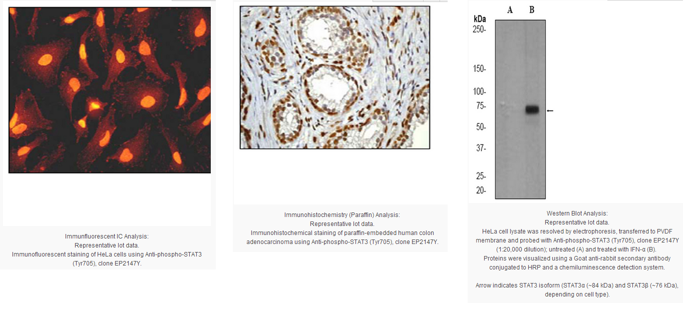
Immunohistochemistry Analysis: A 1:100-250 dilution from a previous lot detected STAT3 in human colon adenocarcinoma tissue.
Immunocytochemistry Analysis: A 1:100-250 dilution from a previous lot detected STAT3 in HeLa cells.
Immunoprecipitation: A 1:40 dilution from a previous lot was used in IP.
Biological Information
Immunogen
Synthetic peptide corresponding to residues surrounding phsophorylated Tyr705 of human STAT3.
Epitope
Phosphorylated Tyr705
Clone
EP2147Y
Host
Rabbit
Specificity
This antibody recognizes STAT3 phsophorylated Tyr705.
The protein encoded by this gene is a member of the STAT protein family. In response to cytokines and growth factors, STAT family members are phosphorylated by the receptor associated kinases, and then form homo- or heterodimers that translocate to the cell nucleus where they act as transcription activators. This protein is activated through phosphorylation in response to various cytokines and growth factors including IFNs, EGF, IL5, IL6, HGF, LIF and BMP2. This protein mediates the expression of a variety of genes in response to cell stimuli, and thus plays a key role in many cellular processes such as cell growth and apoptosis. The small GTPase Rac1 has been shown to bind and regulate the activity of this protein. PIAS3 protein is a specific inhibitor of this protein. Three alternatively spliced transcript variants encodingdistinct isoforms have been described.
FUNCTION:Transcription factor that binds to the interleukin-6 (IL-6)-responsive elements identified in the promoters of various acute-phase protein genes. Activated by IL31 through IL31RA. SUBUNIT STRUCTURE:Forms a homodimer or a heterodimer with a related family member (at least STAT1). Interacts with NCOA1, PELP1, SOCS7 and STATIP1. Interacts with HCV core protein. Interacts with IL23R in presence of IL23. Interacts with IL31RA. Interacts with SIPAR. Interacts (via SH2 domain) with NLK By similarity. Interacts with KPNA4 and KPNA5; KPNA4 may be the primary mediator of nuclear import By similarity. Interacts with TMF1. SUBCELLULAR LOCATION:Cytoplasm. Nucleus. Note: Shuttles between the nucleus and the cytoplasm. Constitutive nuclear presence is independent of tyrosine phosphorylation. TISSUE SPECIFICITY:Heart, brain, placenta, lung, liver, skeletal muscle, kidney and pancreas. PTM:Tyrosine phosphorylated in response to IL-6, IL-11, CNTF, LIF, CSF-1, EGF, PDGF, IFN-alpha and OSM. Phosphorylated on serine upon DNA damage, probably by ATM or ATR. Serine phosphorylation is important for the formation of stable DNA-binding STAT3 homodimers and maximal transcriptional activity. INVOLVEMENT IN DISEASE:Defects in STAT3 are the cause of hyperimmunoglobulin E recurrent infection syndrome autosomal dominant (AD-HIES) [MIM:147060]; also known as hyper-IgE syndrome or Job syndrome. AD-HIES is a rare disorder of immunity and connective tissue characterized by immunodeficiency, chronic eczema, recurrent Staphylococcal infections, increased serum IgE, eosinophilia, distinctive coarse facial appearance, abnormal dentition, hyperextensibility of the joints, and bone fractures. MISCELLANEOUS:Involved in the gp130-mediated signaling pathway. SEQUENCE SIMILARITIES:Belongs to the transcription factor STAT family.
Contains 1 SH2 domain.
Product Usage Statements
Quality Assurance
Evaluated by Western Blot on untreated and IFN-α-treated HeLa cell lysates.
Western Blot Analysis: A 1:10,000-20,000 dilution of this antibody was used to detect STAT3 in IFNα-treated HeLa cell lysate.
Usage Statement
Unless otherwise stated in our catalog or other company documentation accompanying the product(s), our products are intended for research use only and are not to be used for any other purpose, which includes but is not limited to, unauthorized commercial uses, in vitro diagnostic uses, ex vivo or in vivo therapeutic uses or any type of consumption or application to humans or animals.
Storage and Shipping Information
Storage Conditions
Stable for 1 year at -20ºC from date of receipt.
Handling Recommendations: Upon receipt and prior to removing the cap, centrifuge the vial and gently mix the solution. Aliquot into microcentrifuge tubes and store at -20°C. Avoid repeated freeze/thaw cycles, which may damage IgG and affect product performance.
Note: Variability in freezer temperatures below -20°C may cause glycerol containing solutions to become frozen during storage.
STAT proteins (Signal Transduction and Activators of Transcription) are latent cytoplasmic transcription factors that have the dual function of signal transduction and activation of transcription. STATs are activated by tyrosine phosphorylation in response to different ligands, after which they translocate to the cell nucleus. The N-terminal region is highly homologous among the STAT proteins and surrounds a completely conserved arginine residue. STATs are a part of the JAK-STAT signaling pathway – a major pathway of the immune system. All cytokines transduce critical signals through this pathway. STAT3 has been shown to be activated by IFN alpha but not IFN beta. The transcription factors associated with STAT3 are cJun and cyclic AMP responsive enhancer binding protein (CREB). Deletion of the STAT3 gene in knock out mice was lethal at the early embryonic stage.
Product Information
Format
Unpurified
Control
HeLa cell lysate
Presentation
Rabbit Monoclonal in buffer containing 50 mM Tris-Glycine (pH 7.4), 0.15 M NaCl containing 40% Glycerol, 0.01% sodium azide and 0.05% BSA.
Applications
Application
Please note that this product will not be available for sale after March 15, 2015. Please select one of the other antibodies against this target. Anti-phospho-STAT3 (Tyr705) Antibody, clone EP2147Y, Rabbit is an antibody against phospho-STAT3 (Tyr705) for use in WB, IH(P), IC & IP.
Key Applications
Immunoprecipitation
Western Blotting
Immunohistochemistry (Paraffin)
Immunocytochemistry
Application Notes
Immunohistochemistry Analysis: A 1:100-250 dilution from a previous lot detected STAT3 in human colon adenocarcinoma tissue.
Immunocytochemistry Analysis: A 1:100-250 dilution from a previous lot detected STAT3 in HeLa cells.
Immunoprecipitation: A 1:40 dilution from a previous lot was used in IP.
Biological Information
Immunogen
Synthetic peptide corresponding to residues surrounding phsophorylated Tyr705 of human STAT3.
Epitope
Phosphorylated Tyr705
Clone
EP2147Y
Host
Rabbit
Specificity
This antibody recognizes STAT3 phsophorylated Tyr705.
The protein encoded by this gene is a member of the STAT protein family. In response to cytokines and growth factors, STAT family members are phosphorylated by the receptor associated kinases, and then form homo- or heterodimers that translocate to the cell nucleus where they act as transcription activators. This protein is activated through phosphorylation in response to various cytokines and growth factors including IFNs, EGF, IL5, IL6, HGF, LIF and BMP2. This protein mediates the expression of a variety of genes in response to cell stimuli, and thus plays a key role in many cellular processes such as cell growth and apoptosis. The small GTPase Rac1 has been shown to bind and regulate the activity of this protein. PIAS3 protein is a specific inhibitor of this protein. Three alternatively spliced transcript variants encodingdistinct isoforms have been described.
FUNCTION:Transcription factor that binds to the interleukin-6 (IL-6)-responsive elements identified in the promoters of various acute-phase protein genes. Activated by IL31 through IL31RA. SUBUNIT STRUCTURE:Forms a homodimer or a heterodimer with a related family member (at least STAT1). Interacts with NCOA1, PELP1, SOCS7 and STATIP1. Interacts with HCV core protein. Interacts with IL23R in presence of IL23. Interacts with IL31RA. Interacts with SIPAR. Interacts (via SH2 domain) with NLK By similarity. Interacts with KPNA4 and KPNA5; KPNA4 may be the primary mediator of nuclear import By similarity. Interacts with TMF1. SUBCELLULAR LOCATION:Cytoplasm. Nucleus. Note: Shuttles between the nucleus and the cytoplasm. Constitutive nuclear presence is independent of tyrosine phosphorylation. TISSUE SPECIFICITY:Heart, brain, placenta, lung, liver, skeletal muscle, kidney and pancreas. PTM:Tyrosine phosphorylated in response to IL-6, IL-11, CNTF, LIF, CSF-1, EGF, PDGF, IFN-alpha and OSM. Phosphorylated on serine upon DNA damage, probably by ATM or ATR. Serine phosphorylation is important for the formation of stable DNA-binding STAT3 homodimers and maximal transcriptional activity. INVOLVEMENT IN DISEASE:Defects in STAT3 are the cause of hyperimmunoglobulin E recurrent infection syndrome autosomal dominant (AD-HIES) [MIM:147060]; also known as hyper-IgE syndrome or Job syndrome. AD-HIES is a rare disorder of immunity and connective tissue characterized by immunodeficiency, chronic eczema, recurrent Staphylococcal infections, increased serum IgE, eosinophilia, distinctive coarse facial appearance, abnormal dentition, hyperextensibility of the joints, and bone fractures. MISCELLANEOUS:Involved in the gp130-mediated signaling pathway. SEQUENCE SIMILARITIES:Belongs to the transcription factor STAT family.
Contains 1 SH2 domain.
Product Usage Statements
Quality Assurance
Evaluated by Western Blot on untreated and IFN-α-treated HeLa cell lysates.
Western Blot Analysis: A 1:10,000-20,000 dilution of this antibody was used to detect STAT3 in IFNα-treated HeLa cell lysate.
Usage Statement
Unless otherwise stated in our catalog or other company documentation accompanying the product(s), our products are intended for research use only and are not to be used for any other purpose, which includes but is not limited to, unauthorized commercial uses, in vitro diagnostic uses, ex vivo or in vivo therapeutic uses or any type of consumption or application to humans or animals.
Storage and Shipping Information
Storage Conditions
Stable for 1 year at -20ºC from date of receipt.
Handling Recommendations: Upon receipt and prior to removing the cap, centrifuge the vial and gently mix the solution. Aliquot into microcentrifuge tubes and store at -20°C. Avoid repeated freeze/thaw cycles, which may damage IgG and affect product performance.
Note: Variability in freezer temperatures below -20°C may cause glycerol containing solutions to become frozen during storage.


 京公网安备11010802025653 版权所有:北京逸优科技有限公司
