SMAD2 or Mothers against decapentaplegic homolog 2 is a polypeptide that, as its name describes, is a homolog of the Drosophila gene: "Mothers against decepentaplegic". It belongs to the SMAD family of proteins, which belong to the TGFbeta superfamily of modulators. Like many other TGFβ family members SMAD2 is involved in cell signalling. SMAD2 modulates signals of activin and TGFbeta's. It interacts with SMAD anchor for receptor activation (SARA). The binding of ligands causes the phosphorylation of the SMAD2 protein and the dissociation from SARA and the association with SMAD4. It is subsequently transferred to the nucleus where it forms complexes with other proteins and acts as a transcription factor. SMAD2 is a receptor regulated SMAD (R-SMAD) and is activated by bone morphogenetic protein type 1 receptor kinase.
Product Information
Format
Unpurified
Control
Jurkat cell lysate
Presentation
Rabbit Monoclonal in buffer containing 50 mM Tris-Glycine (pH 7.4), 0.15 M NaCl containing 40% Glycerol, 0.01% sodium azide and 0.05% BSA.
Applications
Application
Please note that this product will not be available for sale after March 15, 2015. Please select one of the other antibodies against this target. This Anti-SMAD2 Antibody, clone EP567Y, Rabbit is validated for use in WB, IH(P), IC, FC for the detection of SMAD2.
Key Applications
Flow Cytometry
Western Blotting
Immunohistochemistry (Paraffin)
Immunocytochemistry
Application Notes
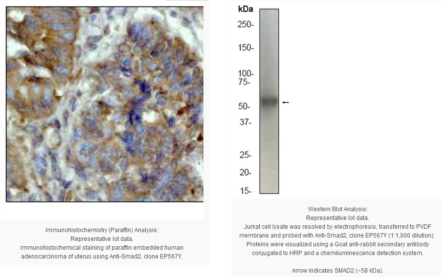
Immunohistochemistry Analysis (paraffin): A 1:50 dilution from a previous lot detected Smad2 in human adenocarcinoma of uterus tissue.
Flow Cytometry: A 1:20 dilution from a previous lot was used in FC.
Immunocytochemistry: A 1:250-500 dilution from a previous lot was used in IC.
Biological Information
Immunogen
Synthetic peptide corresponding to residues surrounding the MH2 domain of human SMAD2.
Epitope
MH2 domain
Clone
EP567Y
Host
Rabbit
Specificity
This antibody recognizes SMAD2 at and around the MH2 domain.
The protein encoded by this gene belongs to the SMAD, a family of proteins similar to the gene products of the Drosophila gene 'mothers against decapentaplegic' (Mad) and the C. elegans gene Sma. SMAD proteins are signal transducers and transcriptional modulators that mediate multiple signaling pathways. This protein mediates the signal of the transforming growth factor (TGF)-beta, and thus regulates multiple cellular processes, such as cell proliferation, apoptosis, and differentiation. This protein is recruited to the TGF-beta receptors through its interaction with the SMAD anchor for receptor activation (SARA) protein. In response to TGF-beta signal, this protein is phosphorylated by the TGF-beta receptors. The phosphorylation induces the dissociation of this protein with SARA and the association with the family member SMAD4. The association with SMAD4 is important for the translocation of this protein into the nucleus, where it binds to target promoters and forms a transcription repressor complex with other cofactors. This protein can also be phosphorylated by activin type 1 receptor kinase, and mediates the signal from the activin. Alternatively spliced transcript variants encoding the same protein have been observed.
FUNCTION:Transcriptional modulator activated by TGF-beta and activin type 1 receptor kinase. SMAD2 is a receptor-regulated SMAD (R-SMAD). May act as a tumor suppressor in colorectal carcinoma. SUBUNIT STRUCTURE:Found in a complex with SMAD3 and TRIM33 upon addition of TGF-beta. Interacts with SMAD3 and TRIM33. Interacts with SARA (SMAD anchor for receptor activation); may form trimers with the SMAD4 co-SMAD. Interacts with FOXH1, homeobox protein TGIF, PEBP2-alpha subunit, CREB-binding protein (CBP), EP300 and SKI. Interacts with SNON; when phosphorylated at Ser-465/467. Interacts (via PY-motif) with SMURF2. Interacts with AIP1 and HGS. Interacts with NEDD4L in response to TGF-beta By similarity. Interacts with LBXCOR1 and CORL2. SUBCELLULAR LOCATION:Cytoplasm. Nucleus. Note: Cytoplasmic in the absence of ligand. Migrates to the nucleus when complexed with SMAD4. TISSUE SPECIFICITY:Expressed at high levels in skeletal muscle, heart and placenta. PTM:Phosphorylated on one or several of Thr-220, Ser-245, Ser-250, and Ser-255. In response to TGF-beta, phosphorylated on Ser-465/467 by TGF-beta and activin type 1 receptor kinases. Able to interact with SMURF2 when phosphorylated on Ser-465/467, recruiting other proteins, such as SNON, for degradation. In response to decorin, the naturally occurring inhibitor of TGF-beta signaling, phosphorylated on Ser-240 by CaMK2. Phosphorylated by MAPK3 upon EGF stimulation; which increases transcriptional activity and stability, and is blocked by calmodulin.
In response to TGF-beta, ubiquitinated by NEDD4L; which promotes its degradation By similarity.
Acetylated on Lys-19 by coactivators in response to TGF-beta signaling, which increases transcriptional activity. Isoform short: Acetylation increases DNA binding activity in vitro and enhances its association with target promoters in vivo. INVOLVEMENT IN DISEASE:Defects in SMAD2 are found in sporadic cases of colorectal carcinoma. SEQUENCE SIMILARITIES:Belongs to the dwarfin/SMAD family.
Contains 1 MH1 (MAD homology 1) domain.
Contains 1 MH2 (MAD homology 2) domain.
Product Usage Statements
Quality Assurance
Evaluated by Western Blot on Jurkat cell lysates.
Western Blot Analysis: A 1:500-1,000 dilution of this antibody was used to detect SMAD2 in Jurkat cell lysate.
Usage Statement
Unless otherwise stated in our catalog or other company documentation accompanying the product(s), our products are intended for research use only and are not to be used for any other purpose, which includes but is not limited to, unauthorized commercial uses, in vitro diagnostic uses, ex vivo or in vivo therapeutic uses or any type of consumption or application to humans or animals.
Storage and Shipping Information
Storage Conditions
Stable for 1 year at -20ºC from date of receipt.
Handling Recommendations: Upon receipt and prior to removing the cap, centrifuge the vial and gently mix the solution. Aliquot into microcentrifuge tubes and store at -20°C. Avoid repeated freeze/thaw cycles, which may damage IgG and affect product performance.
Note: Variability in freezer temperatures below -20°C may cause glycerol containing solutions to become frozen during storage.
SMAD2 or Mothers against decapentaplegic homolog 2 is a polypeptide that, as its name describes, is a homolog of the Drosophila gene: "Mothers against decepentaplegic". It belongs to the SMAD family of proteins, which belong to the TGFbeta superfamily of modulators. Like many other TGFβ family members SMAD2 is involved in cell signalling. SMAD2 modulates signals of activin and TGFbeta's. It interacts with SMAD anchor for receptor activation (SARA). The binding of ligands causes the phosphorylation of the SMAD2 protein and the dissociation from SARA and the association with SMAD4. It is subsequently transferred to the nucleus where it forms complexes with other proteins and acts as a transcription factor. SMAD2 is a receptor regulated SMAD (R-SMAD) and is activated by bone morphogenetic protein type 1 receptor kinase.
Product Information
Format
Unpurified
Control
Jurkat cell lysate
Presentation
Rabbit Monoclonal in buffer containing 50 mM Tris-Glycine (pH 7.4), 0.15 M NaCl containing 40% Glycerol, 0.01% sodium azide and 0.05% BSA.
Applications
Application
Please note that this product will not be available for sale after March 15, 2015. Please select one of the other antibodies against this target. This Anti-SMAD2 Antibody, clone EP567Y, Rabbit is validated for use in WB, IH(P), IC, FC for the detection of SMAD2.
Key Applications
Flow Cytometry
Western Blotting
Immunohistochemistry (Paraffin)
Immunocytochemistry
Application Notes
Immunohistochemistry Analysis (paraffin): A 1:50 dilution from a previous lot detected Smad2 in human adenocarcinoma of uterus tissue.
Flow Cytometry: A 1:20 dilution from a previous lot was used in FC.
Immunocytochemistry: A 1:250-500 dilution from a previous lot was used in IC.
Biological Information
Immunogen
Synthetic peptide corresponding to residues surrounding the MH2 domain of human SMAD2.
Epitope
MH2 domain
Clone
EP567Y
Host
Rabbit
Specificity
This antibody recognizes SMAD2 at and around the MH2 domain.
The protein encoded by this gene belongs to the SMAD, a family of proteins similar to the gene products of the Drosophila gene 'mothers against decapentaplegic' (Mad) and the C. elegans gene Sma. SMAD proteins are signal transducers and transcriptional modulators that mediate multiple signaling pathways. This protein mediates the signal of the transforming growth factor (TGF)-beta, and thus regulates multiple cellular processes, such as cell proliferation, apoptosis, and differentiation. This protein is recruited to the TGF-beta receptors through its interaction with the SMAD anchor for receptor activation (SARA) protein. In response to TGF-beta signal, this protein is phosphorylated by the TGF-beta receptors. The phosphorylation induces the dissociation of this protein with SARA and the association with the family member SMAD4. The association with SMAD4 is important for the translocation of this protein into the nucleus, where it binds to target promoters and forms a transcription repressor complex with other cofactors. This protein can also be phosphorylated by activin type 1 receptor kinase, and mediates the signal from the activin. Alternatively spliced transcript variants encoding the same protein have been observed.
FUNCTION:Transcriptional modulator activated by TGF-beta and activin type 1 receptor kinase. SMAD2 is a receptor-regulated SMAD (R-SMAD). May act as a tumor suppressor in colorectal carcinoma. SUBUNIT STRUCTURE:Found in a complex with SMAD3 and TRIM33 upon addition of TGF-beta. Interacts with SMAD3 and TRIM33. Interacts with SARA (SMAD anchor for receptor activation); may form trimers with the SMAD4 co-SMAD. Interacts with FOXH1, homeobox protein TGIF, PEBP2-alpha subunit, CREB-binding protein (CBP), EP300 and SKI. Interacts with SNON; when phosphorylated at Ser-465/467. Interacts (via PY-motif) with SMURF2. Interacts with AIP1 and HGS. Interacts with NEDD4L in response to TGF-beta By similarity. Interacts with LBXCOR1 and CORL2. SUBCELLULAR LOCATION:Cytoplasm. Nucleus. Note: Cytoplasmic in the absence of ligand. Migrates to the nucleus when complexed with SMAD4. TISSUE SPECIFICITY:Expressed at high levels in skeletal muscle, heart and placenta. PTM:Phosphorylated on one or several of Thr-220, Ser-245, Ser-250, and Ser-255. In response to TGF-beta, phosphorylated on Ser-465/467 by TGF-beta and activin type 1 receptor kinases. Able to interact with SMURF2 when phosphorylated on Ser-465/467, recruiting other proteins, such as SNON, for degradation. In response to decorin, the naturally occurring inhibitor of TGF-beta signaling, phosphorylated on Ser-240 by CaMK2. Phosphorylated by MAPK3 upon EGF stimulation; which increases transcriptional activity and stability, and is blocked by calmodulin.
In response to TGF-beta, ubiquitinated by NEDD4L; which promotes its degradation By similarity.
Acetylated on Lys-19 by coactivators in response to TGF-beta signaling, which increases transcriptional activity. Isoform short: Acetylation increases DNA binding activity in vitro and enhances its association with target promoters in vivo. INVOLVEMENT IN DISEASE:Defects in SMAD2 are found in sporadic cases of colorectal carcinoma. SEQUENCE SIMILARITIES:Belongs to the dwarfin/SMAD family.
Contains 1 MH1 (MAD homology 1) domain.
Contains 1 MH2 (MAD homology 2) domain.
Product Usage Statements
Quality Assurance
Evaluated by Western Blot on Jurkat cell lysates.
Western Blot Analysis: A 1:500-1,000 dilution of this antibody was used to detect SMAD2 in Jurkat cell lysate.
Usage Statement
Unless otherwise stated in our catalog or other company documentation accompanying the product(s), our products are intended for research use only and are not to be used for any other purpose, which includes but is not limited to, unauthorized commercial uses, in vitro diagnostic uses, ex vivo or in vivo therapeutic uses or any type of consumption or application to humans or animals.
Storage and Shipping Information
Storage Conditions
Stable for 1 year at -20ºC from date of receipt.
Handling Recommendations: Upon receipt and prior to removing the cap, centrifuge the vial and gently mix the solution. Aliquot into microcentrifuge tubes and store at -20°C. Avoid repeated freeze/thaw cycles, which may damage IgG and affect product performance.
Note: Variability in freezer temperatures below -20°C may cause glycerol containing solutions to become frozen during storage.


 京公网安备11010802025653 版权所有:北京逸优科技有限公司
