DNA-PK, DNA-protein kinase, is a nuclear protein kinase that recognizes and initiates repair of DNA double strand breaks produced by ionizing radiation and certain drugs.
Product Information
Format
Culture Supernatant
Control
K562 cell lysate
Presentation
Rabbit Monoclonal in buffer containing 50 mM Tris-Glycine (pH 7.4), 0.15 M NaCl containing 40% Glycerol, 0.01% sodium azide and 0.05% BSA.
Applications
Application
Use Anti-PRKDC/DNA-PK Antibody, clone Y393 (rabbit monoclonal antibody) validated in WB,IHC(P), ICC to detect PRKDC/DNA-PK also known as DNA-PK catalytic subunit complementing 1, protein kinase DNA-activated catalytic polypeptide.
Key Applications
Immunocytochemistry
Western Blotting
Immunohistochemistry (Paraffin)
Application Notes
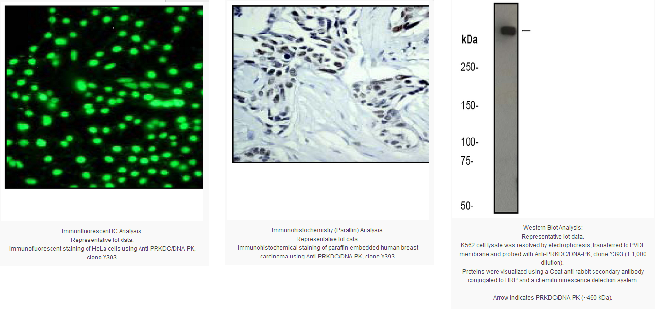
Immunohistochemistry Analysis: A 1:50 dilution from a previous lot detected PRKDC/DNA-PK in human breast carcinoma tissue.
Immunocytochemistry Analysis: A 1:250-500 dilution from a previous lot detected PRKDC/DNA-PK in HeLa cells.
Biological Information
Immunogen
Synthetic peptide corresponding to residues in the C-terminus of human DNA-PK.
Epitope
C-terminus
Clone
Y393
Host
Rabbit
Specificity
This antibody may detect spliced isoform 2 of C-terminal PRKDC/DNA-PK.
The PRKDC gene encodes the catalytic subunit of a nuclear DNA-dependent serine/threonine protein kinase (DNA-PK). The second component is the autoimmune antigen Ku (MIM 152690), which is encoded by the G22P1 gene on chromosome 22q. On its own, the catalytic subunit of DNA-PK is inactive and relies on the G22P1 component to direct it to the DNA and trigger its kinase activity; PRKDC must be bound to DNA to express its catalytic properties.
FUNCTION:Serine/threonine-protein kinase that acts as a molecular sensor for DNA damage. Involved in DNA nonhomologous end joining (NHEJ) required for double-strand break (DSB) repair and V(D)J recombination. Must be bound to DNA to express its catalytic properties. Promotes processing of hairpin DNA structures in V(D)J recombination by activation of the hairpin endonuclease artemis (DCLRE1C). The assembly of the DNA-PK complex at DNA ends is also required for the NHEJ ligation step. Required to protect and align broken ends of DNA. May also act as a scaffold protein to aid the localization of DNA repair proteins to the site of damage. Found at the ends of chromosomes, suggesting a further role in the maintenance of telomeric stability and the prevention of chromosomal end fusion. Also involved in modulation of transcription. Recognizes the substrate consensus sequence [ST]-Q. Phosphorylates 'Ser-139' of histone variant H2AX/H2AFX, thereby regulating DNA damage response mechanism. Phosphorylates DCLRE1C, c-Abl/ABL1, histone H1, HSPCA, c-jun/JUN, p53/TP53, PARP1, POU2F1, DHX9, SRF, XRCC1, XRCC1, XRCC4, XRCC5, XRCC6, WRN, c-myc/MYC and RFA2. Can phosphorylate C1D not only in the presence of linear DNA but also in the presence of supercoiled DNA. Ability to phosphorylate TP53/p53 in the presence of supercoiled DNA is dependent on C1D. CATALYTIC ACTIVITY:ATP + a protein = ADP + a phosphoprotein. ENZYME REGULATION:Inhibited by wortmannin. Activity of the enzyme seems to be attenuated by autophosphorylation. SUBUNIT STRUCTURE:DNA-PK is a heterotrimer of PRKDC and the Ku p70-p86 (XRCC6-XRCC5) dimer. Formation of this complex may be promoted by interaction with ILF3. Associates with the DNA-bound Ku heterodimer, but it can also bind to and be activated by free DNA. Interacts with DNA-PKcs-interacting protein (KIP) with the region upstream the kinase domain. PRKDC alone also interacts with and phosphorylates DCLRE1C, thereby activating the latent endonuclease activity of this protein. Interacts with C1D. SUBCELLULAR LOCATION:Nucleus. PTM:Phosphorylated upon DNA damage, probably by ATM or ATR. Autophosphorylated on Thr-2609, Thr-2638 and Thr-2647. Thr-2609 is a DNA damage-inducible phosphorylation site (inducible with ionizing radiation, IR). Autophosphorylation induces a conformational change that leads to remodeling of the DNA-PK complex, requisite for efficient end processing and DNA repair. SEQUENCE SIMILARITIES:Belongs to the PI3/PI4-kinase family.
Contains 1 FAT domain.
Contains 1 FATC domain.
Contains 2 HEAT repeats.
Contains 1 PI3K/PI4K domain.
Contains 3 TPR repeats.
Product Usage Statements
Quality Assurance
Evaluated by Western Blot on K562 cell lysates.
Western Blot Analysis: A 1:1,000 dilution of this antibody was used to detect PRKDC/DNA-PK in K562 cell lysate.
Usage Statement
Unless otherwise stated in our catalog or other company documentation accompanying the product(s), our products are intended for research use only and are not to be used for any other purpose, which includes but is not limited to, unauthorized commercial uses, in vitro diagnostic uses, ex vivo or in vivo therapeutic uses or any type of consumption or application to humans or animals.
Storage and Shipping Information
Storage Conditions
Stable for 1 year at -20ºC from date of receipt.
Handling Recommendations: Upon receipt and prior to removing the cap, centrifuge the vial and gently mix the solution. Aliquot into microcentrifuge tubes and store at -20°C. Avoid repeated freeze/thaw cycles, which may damage IgG and affect product performance.
Note: Variability in freezer temperatures below -20°C may cause glycerol containing solutions to become frozen during storage.
DNA-PK, DNA-protein kinase, is a nuclear protein kinase that recognizes and initiates repair of DNA double strand breaks produced by ionizing radiation and certain drugs.
Product Information
Format
Culture Supernatant
Control
K562 cell lysate
Presentation
Rabbit Monoclonal in buffer containing 50 mM Tris-Glycine (pH 7.4), 0.15 M NaCl containing 40% Glycerol, 0.01% sodium azide and 0.05% BSA.
Applications
Application
Use Anti-PRKDC/DNA-PK Antibody, clone Y393 (rabbit monoclonal antibody) validated in WB,IHC(P), ICC to detect PRKDC/DNA-PK also known as DNA-PK catalytic subunit complementing 1, protein kinase DNA-activated catalytic polypeptide.
Key Applications
Immunocytochemistry
Western Blotting
Immunohistochemistry (Paraffin)
Application Notes
Immunohistochemistry Analysis: A 1:50 dilution from a previous lot detected PRKDC/DNA-PK in human breast carcinoma tissue.
Immunocytochemistry Analysis: A 1:250-500 dilution from a previous lot detected PRKDC/DNA-PK in HeLa cells.
Biological Information
Immunogen
Synthetic peptide corresponding to residues in the C-terminus of human DNA-PK.
Epitope
C-terminus
Clone
Y393
Host
Rabbit
Specificity
This antibody may detect spliced isoform 2 of C-terminal PRKDC/DNA-PK.
The PRKDC gene encodes the catalytic subunit of a nuclear DNA-dependent serine/threonine protein kinase (DNA-PK). The second component is the autoimmune antigen Ku (MIM 152690), which is encoded by the G22P1 gene on chromosome 22q. On its own, the catalytic subunit of DNA-PK is inactive and relies on the G22P1 component to direct it to the DNA and trigger its kinase activity; PRKDC must be bound to DNA to express its catalytic properties.
FUNCTION:Serine/threonine-protein kinase that acts as a molecular sensor for DNA damage. Involved in DNA nonhomologous end joining (NHEJ) required for double-strand break (DSB) repair and V(D)J recombination. Must be bound to DNA to express its catalytic properties. Promotes processing of hairpin DNA structures in V(D)J recombination by activation of the hairpin endonuclease artemis (DCLRE1C). The assembly of the DNA-PK complex at DNA ends is also required for the NHEJ ligation step. Required to protect and align broken ends of DNA. May also act as a scaffold protein to aid the localization of DNA repair proteins to the site of damage. Found at the ends of chromosomes, suggesting a further role in the maintenance of telomeric stability and the prevention of chromosomal end fusion. Also involved in modulation of transcription. Recognizes the substrate consensus sequence [ST]-Q. Phosphorylates 'Ser-139' of histone variant H2AX/H2AFX, thereby regulating DNA damage response mechanism. Phosphorylates DCLRE1C, c-Abl/ABL1, histone H1, HSPCA, c-jun/JUN, p53/TP53, PARP1, POU2F1, DHX9, SRF, XRCC1, XRCC1, XRCC4, XRCC5, XRCC6, WRN, c-myc/MYC and RFA2. Can phosphorylate C1D not only in the presence of linear DNA but also in the presence of supercoiled DNA. Ability to phosphorylate TP53/p53 in the presence of supercoiled DNA is dependent on C1D. CATALYTIC ACTIVITY:ATP + a protein = ADP + a phosphoprotein. ENZYME REGULATION:Inhibited by wortmannin. Activity of the enzyme seems to be attenuated by autophosphorylation. SUBUNIT STRUCTURE:DNA-PK is a heterotrimer of PRKDC and the Ku p70-p86 (XRCC6-XRCC5) dimer. Formation of this complex may be promoted by interaction with ILF3. Associates with the DNA-bound Ku heterodimer, but it can also bind to and be activated by free DNA. Interacts with DNA-PKcs-interacting protein (KIP) with the region upstream the kinase domain. PRKDC alone also interacts with and phosphorylates DCLRE1C, thereby activating the latent endonuclease activity of this protein. Interacts with C1D. SUBCELLULAR LOCATION:Nucleus. PTM:Phosphorylated upon DNA damage, probably by ATM or ATR. Autophosphorylated on Thr-2609, Thr-2638 and Thr-2647. Thr-2609 is a DNA damage-inducible phosphorylation site (inducible with ionizing radiation, IR). Autophosphorylation induces a conformational change that leads to remodeling of the DNA-PK complex, requisite for efficient end processing and DNA repair. SEQUENCE SIMILARITIES:Belongs to the PI3/PI4-kinase family.
Contains 1 FAT domain.
Contains 1 FATC domain.
Contains 2 HEAT repeats.
Contains 1 PI3K/PI4K domain.
Contains 3 TPR repeats.
Product Usage Statements
Quality Assurance
Evaluated by Western Blot on K562 cell lysates.
Western Blot Analysis: A 1:1,000 dilution of this antibody was used to detect PRKDC/DNA-PK in K562 cell lysate.
Usage Statement
Unless otherwise stated in our catalog or other company documentation accompanying the product(s), our products are intended for research use only and are not to be used for any other purpose, which includes but is not limited to, unauthorized commercial uses, in vitro diagnostic uses, ex vivo or in vivo therapeutic uses or any type of consumption or application to humans or animals.
Storage and Shipping Information
Storage Conditions
Stable for 1 year at -20ºC from date of receipt.
Handling Recommendations: Upon receipt and prior to removing the cap, centrifuge the vial and gently mix the solution. Aliquot into microcentrifuge tubes and store at -20°C. Avoid repeated freeze/thaw cycles, which may damage IgG and affect product performance.
Note: Variability in freezer temperatures below -20°C may cause glycerol containing solutions to become frozen during storage.


 京公网安备11010802025653 版权所有:北京逸优科技有限公司
