Species predicted to react based on 100% sequence homology:Human
Specificity / Sensitivity
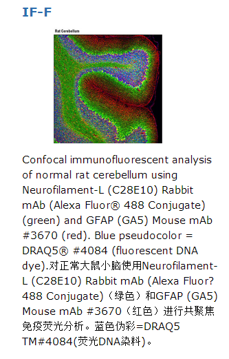
Neurofilament-L (C28E10) Rabbit mAb (Alexa Fluor® 488 Conjugate) detects endogenous levels of total Neurofilament-L protein.
Source / Purification
Monoclonal antibody is produced by immunizing animals with a synthetic peptide surrounding Glu450 of human Neurofilament-L.
Description
This Cell Signaling Technology antibody is conjugated to Alexa Fluor® 488 fluorescent dye and tested in-house for immunofluorescent analysis in human cells. The antibody is expected to exhibit the same species cross-reactivity as the unconjugated Neurofilament-L (C28E10) Rabbit mAb #2837.
Background
The cytoskeleton consists of three types of cytosolic fibers: actin microfilaments, intermediate filaments, and microtubules. Neurofilaments are the major intermediate filaments found in neurons and consist of light (NFL), medium (NFM), and heavy (NFH) subunits (1). Similar in structure to other intermediate filament proteins, neurofilaments have a globular amino-terminal head, a central α-helical rod domain, and a carboxy-terminal tail. A heterotetrameric unit (NFL-NFM and NFL-NFH) forms a protofilament, with eight protofilaments comprising the typical 10 nm intermediate filament (2). While neurofilaments are critical for radial axon growth and determine axon caliber, microtubules are involved in axon elongation. PKA phosphorylates the head domain of NFL and NFM to inhibit neurofilament assembly (3,4). Neurofilament accumulations are found in many human neurological disorders including Parkinson's disease (in Lewy bodies along with α-synuclein), Alzheimer's disease, Charcot-Marie-Tooth disease, and Amyotrophic Lateral Sclerosis (ALS) (1).


 京公网安备11010802025653 版权所有:北京逸优科技有限公司
