来自Cell Signaling Technology (CST)的SignalSilence®LC3B siRNA I 可以帮助研究者通过RNA干扰特异性地抑制 LC3B 的表达,这种方法可以通过将双链RNA分子传递到细胞内从而使基因表达有选择的沉默。LC3B siRNA I 不能抑制LC3A或LC3C的表达。来自CST的所有的SignalSilence®siRNA产品都是经过内部严格检测的,并且通过Western blot 分析证明确实能够减少目的蛋白的表达。通过三苯甲基分析每个碱基以监测寡核苷酸的合成,确保合适的配对效率。随后寡核苷酸通过亲和固相萃取法纯化。退火的RNA双链通过质谱分析来证实其精确的组成。每一批产品都通过质谱分析与前面的产品进行比较,来保证不同批次之间的最大一致性。CST推荐使用100 nM SignalSilence®LC3B siRNA I 进行转染,48到72小时后对细胞进行裂解。转染步骤按照转染试剂说明书提供的步骤进行。遇到任何使用方面的问题,请随时联系CST。自噬是自噬溶酶体对其包裹的细胞质内含物进行降解的一种分解代谢过程(1,2)。自噬过程一般是在营养匮乏的条件下被激活,但是也与一些生理过程有关,如发育、分化、神经变性、感染和癌症(3)。自噬微管相关蛋白的轻链3(LC3)最初被认定为是微管相关蛋白1A和1B的一个亚基(被称为MAP1LC3)(4),随后发现该蛋白所包含的与酵母蛋白类似的Apg8/Aut7/Cvt5对于自噬起着至关重要的作用(5)。三种人类LC3亚型(LC3A、LC3B和LC3C)在自噬过程中翻译后被修饰(6-9)。随着合成后LC3羧基末端迅速裂解,产生了胞浆的LC3-I 型。在自噬过程中,通过一个涉及Atg7和Agt3的类泛素体系,LC3-I被脂质化为LC3-II,从而将LC3与自噬小泡联系起来(6-10)。自噬体中LC3的存在,及其向低迁移形式的LC3-II的转化被作为自噬发生的“指示器”(11)。
SignalSilence® LC3B siRNA I from Cell Signaling Technology (CST) allows the researcher to specifically inhibit LC3B expression using RNA interference, a method whereby gene expression can be selectively silenced through the delivery of double stranded RNA molecules into the cell. LC3B siRNA I will not inhibit expression of LC3A or LC3C. All SignalSilence® siRNA products from CST are rigorously tested in-house and have been shown to reduce target protein expression by western analysis.
Quality Control
Oligonucleotide synthesis is monitored base by base through trityl analysis to ensure appropriate coupling efficiency. The oligo is subsequently purified by affinity-solid phase extraction. The annealed RNA duplex is further analyzed by mass spectrometry to verify the exact composition of the duplex. Each lot is compared to the previous lot by mass spectrometry to ensure maximum lot-to-lot consistency.
Directions for Use
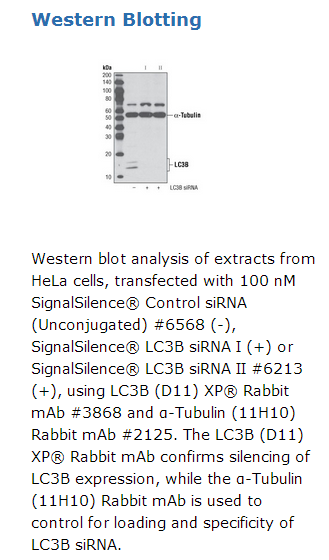
CST recommends transfection with 100 nM LC3B siRNA I 48 to 72 hours prior to cell lysis. For transfection procedure, follow protocol provided by the transfection reagent manufacturer. Please feel free to contact CST with any questions on use.
Background
Autophagy is a catabolic process for the autophagosomic-lysosomal degradation of bulk cytoplasmic contents (1,2). Autophagy is generally activated by conditions of nutrient deprivation but has also been associated with a number of physiological processes including development, differentiation, neurodegenerative diseases, infection and cancer (3). Autophagy marker Light Chain 3 (LC3) was originally identified as a subunit of microtubule-associated proteins 1A and 1B (termed MAP1LC3) (4), and subsequently found to contain similarity to the yeast protein Apg8/Aut7/Cvt5 critical for autophagy (5). Three human LC3 isoforms (LC3A, LC3B, and LC3C) undergo post-translational modifications during autophagy (6-9). Cleavage of LC3 at the carboxy terminus immediately following synthesis yields the cytosolic LC3-I form. During autophagy, LC3-I is converted to LC3-II through lipidation by a ubiquitin-like system involving Atg7 and Atg3 that allows for LC3 to become associated with autophagic vesicles (6-10). The presence of LC3 in autophagosomes and the conversion of LC3 to the lower migrating form LC3-II have been used as indicators of autophagy (11).


 京公网安备11010802025653 版权所有:北京逸优科技有限公司
