Phospho-Talin (Ser425)抗体仅能识别425位丝氨酸磷酸化后的内源性talin蛋白。合成对应人talin蛋白425位丝氨酸及其邻近氨基酸残基序列的多肽免疫动物获得单克隆抗体。抗体由蛋白A和肽亲和层析纯化获得。粘着斑连接细胞骨架与细胞外基质(ECM),一种围绕在哺乳动物器官和组织外由分泌型大分子构成的复杂结构。整联蛋白簇生在粘着斑细胞外侧,传递细胞外基质信号进入细胞内蛋白复合物,随后传导肌动蛋白丝细胞骨架信号以调节张力适应细胞运动。内部信号也可以在粘着斑汇集以调节整联蛋白的亲和性和亲和力。通过粘着斑的信号调节细胞粘附,迁移,扩增,凋亡和基因表达,并影响细胞生命过程包括发育,损伤修复,ianyifanying,侵润,转移和血管生成(reviewed in 1-3)。Talin是一种大型,多结构域粘着斑蛋白,与正脸蛋白和其他粘着蛋白细胞内结构域结合。Talin涉及到粘着斑形成,并将粘着斑与肌动蛋白丝细胞骨架连接(4)。Talin和整合蛋白的结合会提高整合蛋白及ECM中不可溶和可溶蛋白的亲和性(5,6)。使用质谱分析talin245位丝氨酸磷酸化。该位点是CDK5的潜在靶点(7)。
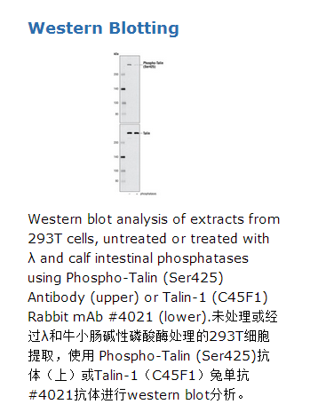
Phospho-Talin (Ser425) Antibody detects endogenous levels of talin protein only when phosphorylated at Ser425.
Source / Purification
Polyclonal antibodies are produced by immunizing animals with a synthetic peptide corresponding to residues surrounding Ser425 of human talin protein. Antibodies are purified using protein A and peptide affinity chromatography.
Background
Focal adhesions connect the cytoskeleton with the extracellular matrix (ECM), a complex structure of secreted macromolecules that surrounds mammalian organs and tissues. Integrins clustered on the extracellular side of focal adhesions signal from the ECM to intracellular protein complexes, which in turn signal to the actin cytoskeleton to regulate the tension needed for cell motility. Internal signals also converge on focal adhesions to regulate integrin affinity and avidity. Signaling through focal adhesions regulates cell adhesion, migration, proliferation, apoptosis and gene expression, and impacts cellular processes such as development, wound healing, immune response, invasion, metastasis and angiogenesis (reviewed in 1-3). Talin is a large, multidomain focal adhesion protein that interacts with the intracellular domains of integrins and other focal adhesion proteins. Talin is involved in the formation of focal adhesions and in linking focal adhesions to the actin cytoskeleton (4). The interaction between talin and integrins increases the affinity between integrin and both insoluble and soluble ECM proteins (5,6).Phosphorylation of talin at Ser425 was discovered using mass spectrometry. This site is a potential substrate of CDK5 (7).


 京公网安备11010802025653 版权所有:北京逸优科技有限公司
