Alexa Fluor®和其他许多阴离子荧光染料和蛋白质可以非特异性地结合阳离子细胞和组织组分。通过有效地阻断这些非特异的静电作用,Image-iT® FX Signal Enhancer能够大大改善免疫标记的细胞和组织的信噪比。Image-iT®是一种液体,在用荧光探测剂染色前,可直接用于有固定的可渗透化处理的细胞或组织样品的载玻片或者盖玻片。
样品准备:1.用透化缓冲液进行细胞的可渗透化处理,室温下作用5分钟。2.用PBS清洗3次,每次5分钟。3.滴加3-4滴Image-iT™ FX Signal Enhancer(或能够覆盖细胞的体积),在室温下孵育30分钟。4. 用PBS清洗3次,每次5分钟。5.用封闭液封闭样品,作用60分钟。6.在封闭的同时准备一抗,用抗体稀释缓冲液按照数据表上的指示进行稀释。7.吸出封闭液,加入稀释好的一抗。8.4°C孵育过夜。9.用PBS清洗3次,每次5分钟。10.盖玻片上滴加Prolong® Gold Antifade Reagent。11.为了获得最好的结果,样品立即用合适的激发波长检测。要长期保存,将载玻片平放,避光保存在4°C。
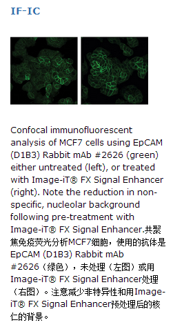
原厂资料:
Alexa Fluor® and many other anionic fluorescent dyes and proteins can bind nonspecifically with cationic cell and tissue constituents. By efficiently blocking these nonspecific electrostatic interactions, Image-iT® FX Signal Enhancer can dramatically improve the signal-to-noise ratio of immunolabeled cells and tissues. Image-iT® is a liquid that is applied directly to slides or coverslips containing fixed and permeabilized cell or tissue samples prior to staining with fluorescent probes.
Directions for Use
Following specimen preparation: 1. Permeabilize the cells in Permeabilization Buffer for 5 minutes at room temperature. 2. Rinse three times in PBS for 5 minutes each. 3. Apply 3–4 drops of Image-iT™ FX Signal Enhancer (or sufficient volume to cover the cells) and incubate for 30 minutes at room temperature. 4. Rinse three times with PBS for 5 minutes each. 5. Block specimen in Blocking Buffer for 60 minutes. 6. While blocking, prepare primary antibody by diluting as indicated on datasheet in Antibody Dilution Buffer. 7. Aspirate blocking solution, apply diluted primary antibody. 8. Incubate overnight at 4°C. 9. Rinse three times in PBS for 5 minutes each. 10. Coverslip slides with Prolong® Gold Antifade Reagent. 11. For best results, examine specimens immediately using appropriate excitation wavelength. For long-term storage, store slides flat at 4°C protected from light.
注意事项:
Storage: Store at room temperature. This product is stable for 12 months.


 京公网安备11010802025653 版权所有:北京逸优科技有限公司
