收藏本站
热门搜索: A3500  E1910  A1360  25200056  dmem  

H2AX (pS139) Alexa 555 N1-431 100Tst

  • 产品编号:BD-560446      品牌:BD-Pharmingen       原厂货号:560446
  • 产品分类:抗体 > 一抗 > 磷酸化抗体
  • 应用分类:
 
包装: 100Tst
运保温度: Store undiluted at 4°C
到货周期: 登录后查看
标准价: ¥客户可见
会员价: ¥客户可见
积分: 客户可见
 
登录之后可加入购物车购买,请您 
对比 收藏

运费与支付说明:

1.含干冰类产品有运费;

2. 必须现金付款或有信用额度的会员才可以直接发货,否则需要等待现金付款信息。

描述:

Histones are highly basic proteins that complex with DNA to form chromatin. The H2AX histone (~15 kDa calculated molecular weight) is a
member of the H2A histone family whose members are components of nucleosomal histone octamers. Double-stranded breaks in DNA caused by
replication errors, apoptosis, or other physiological processes (including, immunoglobulin and TCR gene recombinations) and DNA damage caused by
ionizing radiation, UV light, or cytotoxic agents lead to phosphorylation of H2AX on serine 139. H2AX (pS139) is also referred to as H2AX
(pS140) when the N-terminal methionine that is normally excised during posttranslational processing is included in amino acid sequence numbering.
Kinases such as ataxia telangiectasia mutated (ATM) or ATM-Rad3-related (ATR) phosphorylate H2AX to induce its function. Phosphorylated
H2AX (also termed, gamma-H2AX) functions to recruit and localize DNA repair proteins or cell cycle checkpoint factors to the DNA-damaged
sites. In this way, phosphorylated H2AX promotes DNA repair and maintains genomic stability and thus helps prevent oncogenic transformations.
Immunofluorescent staining and bioimaging analysis of cultured cells can be used to readily identify H2AX (pS139)-containing foci. As such, H2AX
(pS139) immunofluorescence localization serves as a biomarker for nuclear sites of DNA damage (e.g., double-stranded DNA breaks) in affected cells.
Immunofluorescent staining of human cell line. HeLa cells (ATCC CCL-2) were seeded in a BD Falcon™ 96-well Imaging
Plate (Cat. No. 353219) at ~10,000 cells per well. After overnight culture, the cells were exposed to 2400 Joules UV
irradiation (right image) or untreated (left image) and then allowed to recover for 30-60 minutes at 37°C. The cells were fixed,
permeabilized with cold methanol, and stained with Alexa Fluor® 555 Mouse anti-H2AX (pS139) (pseudo colored red) according
to the Recommended Assay Procedure. Cell nuclei were counterstained with Hoechst 33342 (pseudo colored blue). The
images were captured on a BD Pathway™ 435 high-content Bioimager system using a 20X objective and merged using BD
AttoVision™ software. This antibody also worked with the Saponin and the Triton X-100 Perm/Wash protocols (see
Recommended Assay Procedure; Bioimaging protocol link).
Preparation and


注意事项:


1.Since applications vary, each investigator should titrate the reagent to obtain optimal results.
2.Caution: Sodium azide yields highly toxic hydrazoic acid under acidic conditions. Dilute azide compounds in running water before discarding to avoid accumulation of potentially explosive deposits in plumbing. .
3.Store undiluted at 4°C and protected from prolonged exposure to light. Do not freeze.
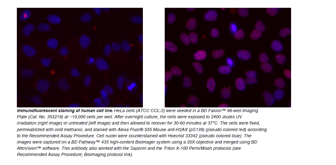

相关图片

本产品可用于的实验

购此产品的人还购买了
最近浏览产品

京ICP备15036693号-2    京公网安备11010802025653    版权所有:北京逸优科技有限公司       0.11

 纠 错