Description: The eBioOMAK-D antibody reacts with mouse perforin (pore-forming protein, pfp, Prf). Perforin is one of the cytolytic mediators present in the cytoplasmic granules of cytotoxic T lymphocytes (CTL) and natural killer cells (NK). Perforin is involved in the killing function by CTLs and NKs and has an important role in the immune response against tumors and virus infections.
By immunobloting, eBioOMAK-D recognizes a ~70kDa band in lysates of CTLL-2 mouse cytotoxic cell line and in lysates of IL-2 stimulated but not unstimulated mouse splenocytes. By multi-color intracellular flow cytometric analysis, eBioOMAK-D staining is increased upon stimulation (IL-2 or anti-CD3/28). Intracellular flow staining results showing upregulation of protein expression have been confirmed by immunoblotting. Furthermore, stimulated Perforin Knock-out (developed by Walsh) splenocytes do not stain with eBioOMAK-D nor is any protein detectable by western blotting with eBioOMAK-D as well as other anti-mouse perforin antibodies. Please note that the Kagi perforin knock-out mice may synthesize a truncated form of the protein which may be recognized by eBioOMAK-D.
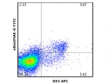
In IL-2 stimulated mouse splenocytes, NK cells (as determined by CD49b staining) contain perforin while CD8 cells contain little to none and can vary with culture conditions. This has been confirmed by staining and western blotting the two populations using both OMAK-D and P1-8 antibodies. In contrast stimulation of splenocytes with anti-CD3/CD28 antibodies does result in an increase of perforin on both NK cells and CD8 cells.
eBioOMAK-D is also crossreactive to human perforin and co-stains CD56 positive cells in PBMC.
Expression of perforin and Granzyme B do not always correlate (as discussed above in the CD8 population of IL-2 stimulated splenocytes). Granzyme B typically is expressed earlier and at higher levels. Expression of Granzyme B is dramatically increased (more than 10,00 fold based on mRNA estimates and significantly at the protein level based on western blotting and flow analysis) compared to a minimal increase (10-100 fold) in perforin mRNA and protein with IL-2 stimulation.
For intracellular staining and flow cytometric analysis with direct conjugates of anti-mouse perforin, it is highly recommended to use the Foxp3 buffer system (cat. 00-5523). Other buffers may yield varying results. For more information, please contact technical support at tech@ebioscience.com.


 京公网安备11010802025653 版权所有:北京逸优科技有限公司
